Online Purchasing Account You are logged on as Guest. LoginRegister a New AccountShopping cart (Empty)
United States 

Cancer

Integrated Solutions for Basic Research
& Translational Discovery

Cancer

Cancer is the leading cause of death worldwide, accounting for nearly one in six deaths. In order to navigate the complex landscape of mutations and diverse types of genetic aberration that leads to cancer, the hallmarks of cancer were proposed as a set of functional capabilities acquired by human cells as normal cells transform into tumor cells in a multistage process that generally progresses from pre-cancerous lesion to a malignant tumor.

As cancer hallmarks have evolved over the last two decades so has our portfolio of tools to support cancer research. Over this time, we have called upon our diverse scientific and technical expertise to create innovative technologies, assays and reagents to aid with genomics, protein, cellular and tissue analysis, in addition to small molecules chemistry.

With our comprehensive catalog of unique products, we are committed to providing the tools and support you need to drive innovation and advancement in cancer and help discover the next hallmark.


Unraveling the Complex Landscape of Cancer

Target Identification

Whether it's a specific gene, protein, lipids or a whole pathway, target identification is one of the most critical steps in cancer research and drug discovery.

Target Characterization

Some targets can play different roles depending on the cell type or microenvironment, hence, the characterization step is crucial for the understanding of the functional role that plays in normal cells vs cancer cells.

Target Analysis

Validation of the target is achieved with target analysis to understand and control its regulatory mechanism and the role that plays in the hallmarks of cancer.

Innovation to Meet the Needs of Evolving Cancer Hallmarks

Target Identification

Whether it's a specific gene, protein, lipids or a whole pathway, target identification is one of the most critical steps in cancer research and drug discovery.


AMPIVIEW™ RNA Probes

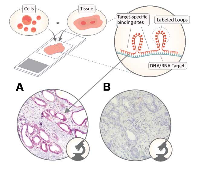
AMPIVIEW™ RNA probes are uniquely designed with the precision of targeted, sequence-specific RNA, powered by Enzo’s LoopRNA™ ISH technology to deliver superior sensitivity when combined with Enzo’s immunochemistry detection solutions. With sample morphology preserved, the spatial localization of target biomarkers can be visualized under the light microscope.

  • Detection of unique nucleic acid targets (DNA/RNA or RNA) down to a single cell level
  • High sensitivity and specificity
  • Adaptable products ready for any workflow (Manual or Automated)


Related Products
AMPIVIEW™ EGFR (AS) Dig RNA Probes Set
AMPIVIEW™ HPV 6/11 RNA Probes Set
AMPIVIEW™ HPV 16/18 RNA Probes Set
AMPIVIEW™ HPV 31/33/51 RNA Probes Set
AMPIVIEW™ HPV High-Risk RNA Probes Set
AMPIVIEW™ Ubiquitin and NSP Dig Control Probes Kit
AMPIVIEW™ GAPDH and NSP Dig Control Probes Kit

AMPIVIEW™ RNA Probes Deliver Superior Sensitivity and Specificity

AMPIVIEW™ RNA probes workflow diagram. Detection of epidermal growth factor receptor (EGFR) in prostate tissue with A. AMPIVIEW™ EGFR (AS) Dig RNA probes and B. AMPIVIEW™ NSP Dig RNA probes, amplified with DIGX® anti-digoxigenin, detected with POLYVIEW® PLUS AP combined with HIGHDEF® Red AP chromogen and hematoxylin.


Chromogenic in situ Hybridization E-book

  • An Introduction to Chromogenic in situ Hybridization (CISH)
  • Top 10 Tips for Successful CISH
  • Methods to Generate Probes
  • Biotin or Digoxigenin
  • DNA vs RNA Probes
  • Sample Preparation
  • Hybridization
  • Controlling Non-specific Background with Blocking
  • Methods of Detection for CISH
  • Single vs Dual Staining
  • Common Counterstaining for CISH
  • Applications of CISH
  • FAQs on CISH

Genome instability and mutations are two of the most common causes of cancer. Fluorescence in situ hybridization (FISH) techniques provides a way to visualize and map the genetic material in individual cells. This is a very useful tool to understand a variety of chromosomal abnormalities and other genetic mutations.


DEEPSEE® CDKN2A/CEN3/7/17 Quad Probe Assay

Ready-to-use probes, used to detect the most frequently encountered chromosomal abnormalities in bladder cancer, contain fluorescently labeled nucleic acid targeting CDKN2A or p16 (gold) and three chromosomes, 3 (red), 7 (green) and 17 (aqua).

  • Validated for use with cells and urine samples
  • Ready-to-use probe mix to minimize pipetting errors
  • Rapid detection of chromosomal aneuploidies and identification of loss


Related Products
CYTAG® CGH Labeling Kit
CYTAG® SuperCGH Labeling Kit
CYTAG® Total CGH Labeling Kit
CD3ε Recombinant Monoclonal Antibody
CD4 Recombinant Monoclonal Antibody
CD19 Recombinant Monoclonal Antibody
CD23 (Human) Monoclonal Antibody
CD33 Recombinant Monoclonal Antibody
CD44std (Human) Monoclonal Antibody

Quadruple Color Assay for the Detection of CDKN2A/CEN3/7/17

A. Normal cytological specimen hybridized with DEEPSEE® CDKN2A/CEN3/7/17 Quad Probe. Two red (CEN 3), two green (CEN 7), two aqua (CEN 17), and two gold (CDKN2A) signals indicating normal interphase nucleus. B. Right: UM-UC-3 bladder cancer cells hybridized with DEEPSEE® CDKN2A/CEN3/7/17 Quad Probe showing trisomy 9, indicated by 3 CDKN2A (gold) signals in the nucleus.


Target Characterization

Some targets can play different roles depending on the cell type or microenvironment, hence, the characterization step is crucial for the understanding of the functional role that plays in normal cells vs cancer cells.


ROS-ID® Hypoxia/Oxidative Stress Detection Kit

ROS-ID® Hypoxia/Oxidative Stress Detection Kit is designed for functional detection of hypoxia and oxidative stress levels in live cells (both suspension and adherent) using fluorescent microscopy or flow cytometry.

  • Detection of hypoxia with high sensitivity, selectivity and accuracy in cacer tissues and cells
  • Allow for simple single-step detectin of hypoxia in non-fixed cells and tissues
  • Dyes can be combined with ROS detection reagents for multiplexing


Related Products
ROS-ID® Total ROS/Superoxide Detection Kit
GFP-CERTIFIED® Apoptosis/Necrosis Detection
NUCLEAR-ID® Green Cell Cycle Kit
PKA Kinase Activity Kit
Akt Kinase Activity Kit
PKC Kinase Activity Kit

Multiplex Hypoxia and Oxidative Stress Detection

HeLa cells were seeded on microscope slides and next day treated with DFO (chemical inducer of hypoxia) or pyocyanin (oxidative stress inducer) for 4 h at 37°C as described in the manual. ROS-ID® was used to detect hypoxia (red) and oxidative stress (green). Post-treatment, slides were washed with PBS, cover-slipped and visualized using an Olympus BX-51 fl uorescence microscope.


Understanding the roles of DNA damage repairs in the promotion of cancer is essential for the development of targeted cancer therapy.

Exposure of cells to oxidative and environmental stresses frequently results in the breakdown of genomic DNA.


DNA Damage ELISA Kit

The DNA Damage ELISA kit is a colometric competitive enzyme immnoassay kit that evaluates the integrity of genomic DNA by assessing the presence of oxidized DNA that are frequently used to verify the onset of apoptosis or DNA damage.

Rapid DNA Damage ELISA kit for cancer, apoptosis and oxidative stress.

  • Reliable and validated in a variety of sample matrices
  • Fully quantitative results that surpass semi-quantitative Western blot analysis
  • No edge effect allowing to assay more samples per kit


Related Products
IL-8 (human), ELISA Kit
IL-33 (human) ELISA Kit
VEGF (human) ELISA Kit
cAMP complete ELISA Kit
cGMP complete ELISA Kit

Highly Sensitive DNA Damage Measurements for Cancer, Apoptosis and Oxidative Stress

Standard Curve of DNA damage. Highly sensitive assay, detecting as low as 1ng/ml with results in less than 2.5 hours.


Target Analysis

Validation of the target is achieved with target analysis to understand and control its regulatory mechanism and the role that plays in the hallmarks of cancer.

Programmed cell death, such as apoptosis and autophagy, are regulated by a variety of biomacromolecures. Emerging evidence has revealed that programmed cell death are key features of tumorigenesis.


CYTO-ID® Autophagy Detection Kit 2.0

Monitor autophagy in live cells with CYTO-ID® Autophagy Detection Kit 2.0, which contains a brighter and more photostable dye that stains autophagic vesicles specifically.

  • Rapid, no transfection required and only 30 minutes of incubation
  • Negligible staining of lysosomes reduces background seen with other dyes
  • Facilitates high-throughput screening of activators and inhibitors of autophagy


Related Products
SCREEN-WELL® Cancer Library
SCREEN-WELL® Epigenetic Library
SCREEN-WELL® FDA Approved Drug Library
27-Hydroxycholesterol
Afatinib
(R)-BI-2536
Ko-143

Quantify Autophagic Vacuoles Without Transfection

CYTO-ID® Autophagy Detection Kit 2.0 (ENZ-KIT175) used to detect autophagy in HeLa cells cultured in (A) media under normal conditions, (B) starvation media (EBSS) treated with 40uM Chloroquine for 4 hours. Starved cells show a higher quantity of autophagic vacuoles compared to cells under normal conditions (fluorescent green).


High quality biomarkers for cancer, especially antibodies, are crucial tools for prevention, diagnosis, prognostics, therapy and to effectively monitor treatment response after disease modifying treatments.

Enzo's portfolio of antibodies for cancer research includes thousands of monoclonal and polyclonal antibodies validated for various applications, including immunohistochemistry (IHC), immunofluorescence (IF), flow cytometry and Western blotting.


High Quality Antibodies for Cancer Research

Enzo offers an extensive repertoire of high-specificity antibodies for the study of cancer hallmarks, including predictive biomarkers such as p53, EGFR or p16.

  • Rigorously validated in serveal applications
  • Superb perfomance documented in thousands of peer-reviewed publications
  • Backed by Enzo's Worry-free Antibody Trial Program


Related Products
POLYVIEW® PLUS HRP (anti-rabbit) Reagent
POLYVIEW® PLUS AP (anti-rabbit) Reagent
POLYVIEW® PLUS HRP (anti-mouse) Reagent
POLYVIEW® PLUS AP (anti-mouse) Reagent
HIGHDEF® DAB Chromogen/Substrate Set
HIGHDEF® Green AP Chromogen/Substrate Set
HIGHDEF® Hematoxylin

Highly Sensitive and Specific Detection of p53 in FFPE Tissue

p53 detection endometrial carcinoma with p53 monoclonal antibody (PAb122)

Search for Antibodies


Antibodies Related to Cancer
p16, p53, Ki67, LAG-3, HER-2, EGFR, Cytokeratin 1/5/10/14, p504S p63 + HMWCK cocktail, PD-1, PD-L1, CTLA-4
 

IHC E-book

  • Top 10 Tips for Successful Immunohistochemistry
  • Choosing your Fixation, Embedding, and Antigen Retrieval Method for Successful IHC
  • Controlling Unspecific Background with Blocking
  • Selecting and Optimizing Primary Antibodies for IHC
  • How to Select a Secondary Antibody
  • Various Detection Methods for IHC
  • Choosing the Right Reporter Enzyme and Chromogen
  • Multiplexing Chromogenic IHC
  • Common Counterstains for IHC
  • Useful Controls for Immunohistochemistry
  • IHC Art Gallery
  • Immunohistochemistry Troubleshooting Guide

Enabling Cancer Research

Leverage over 45 years of innovation and technical expertise supporting drug development, discovery, research, and diagnostics through our core technology platforms. With over 150,000 citations, GMP and ISO certifications, our highly-sensitive, quality products consistently deliver trusted results. We maintain product integrity and reliability with our in-house U.S. manufacturing facility. We deploy our bench of expert PhD scientists to provide technical, tailored, support for our customers and their projects.

Rely on our extensive experience in innovation, technology development, and manufacturing to support your cancer research and development. Our comprehensive Life Sciences Contract Services support the development of customizable, unique, and efficient solutions for all your research needs.

Our Technological Depth Enabling Efficiency for our Customers.


Fighting Cancer with the Immune System

Immuno-oncology, also known as cancer immunotherapy, refers to the use of a person’s own immune system for the treatment of cancer. Research in the field includes:

  • Targeting immune tolerance via co-inhibitory immune checkpoints
  • Enhancing immune responses by costimulatory checkpoint pathways
At Enzo, our diverse scientific and technical expertise has helped us create innovative assays and reagents to help support cancer immunotherapy. We offer a complete set of solutions for Immuno-oncology research including antibodies, ELISA kits and recombinant proteins.


Akt Pathway Discovery

From Target Selection to Lead Profiling

Akt/PKB has emerged as a central player in the signal transduction pathways activated in response to growth factors or insulin. It is thought to contribute to several cellular functions including cell growth, transcriptional regulation, cell survival and nutrient metabolism. It has been linked to a range of diseases including cancer and type II diabetes.

The Akt/PKB pathway is a popular target since it sits at the crossroads of both oncogenic and tumor suppressor signaling pathways. Enzo provides novel tools to help support cancer discovery from target selection, hit-to-lead discovery, and lead profiling.


Serine Proteases and Cancer

Novel Serine Protease Cancer Biomarker Products

Enzo Life Sciences offers more than 40 unique serine protease related products specifically related to cancer biomarker discovery, with emphasis on key family members identified as markers of early stage ovarian, breast, prostate and lung cancer.

  • High-sensitivity, easy-to use and rapid ELISA kits
  • Ready-to-user proteins for use as ELISA or Western blot standards
  • Exclusive antibodies created against native proteins


Related Documents

Latest Literature

Brochures
Cancer
 

Recommend this page