Immunohistochemistry (IHC) is a routinely used technique that uses antibodies to detect the presence and location of proteins and other antigens in tissue sections. Irrespective of detection method (direct or indirect), the antibody-antigen interaction is then visualized using a chromogenic method or a fluorescent-based method. In the direct detection method, the primary antibody is directly label with a reporter. The primary antibody can be labeled with either a reporter enzyme to react with appropriate color-developing substrate or a fluorescent reporter molecule to generate a signal that tells you the antigens’ presence in a tissue section upon binding. The indirect detection method, instead has a secondary antibody directly label to a reporter, with the intention of binding to an unlabeled primary antibody to amplify the initial detection signal. The labeled secondary antibody is raised against the host species and antibody type of the primary antibody. This amplification is possible through activation of the conjugated reporter enzyme or fluorescent molecule to the secondary antibody after the secondary antibody has bound to a single primary antibody targeting the antigen of interest.
During the planning steps of an IHC assay, end-users will have to decide between which approach to use, indirect or direct, which is dependent upon the expected expression levels of the antigen of interest. Indirect detection is suitable for all quantities of antigen expression. This flexibility in sensitivity is provided by the secondary antibodies and what they are conjugated to, in order to amplify signal to detect various quantities of antigens of interest. Direct detection is best suited for studying highly expressed antigens, used when amplification of the signal through a secondary antibody might not be necessary. A major advantage is that direct detection methods offer flexibility with the design of multi-color fluorescent staining experiments that are possible through the various different fluorescent molecules that can be conjugated to the primary antibody. This does not limit an end-user based on the availability of different primary antibodies which can sometimes restricted to only being able to use a single one.
Additionally, prior to running IHC, in antibody-mediated antigen detection, the reporter being selected is a major decision that needs to be considered to visually identify the target antigen. This is based on the type of experiment, the level of antigen expression (as discussed above), whether or not signal quantitation is required, the kinds of imaging needed and the cost of reagents and equipment. As we previously mentioned, irrespective of the detection method being direct or indirect, the most popular methods of detection are enzyme and fluorescent-mediated detection. Fluorophore-based detection with tissue sections is really immunofluorescence (IF) detection and can provide a higher signal-to-noise ratio than chromogenic labels and are thus, more frequently used in simultaneous detection of multiple targets. The scope of this article will focus on chromogenic detection and elaborate further on two commonly used reporter enzymes, Horseradish Peroxidase (HRP) and Alkaline Phosphatase (AP) to discuss the differences to consider when choosing between substrate/chromogens for HRP & AP in your IHC experiments.
Horseradish Peroxidase (HRP)
What are some chromogens? What are some considerations?
Horseradish Peroxidase (HRP) is a 44-kDa protein that catalyzes the oxidation of substrates once activated in the presence of hydrogen peroxide (H
2O
2). Activated HRP oxidizes electron donors such as substituted phenylenediamines and MBTH converting them to cationic electrophiles which are then reacted with electron-rich, aromatic compounds to yield a colored product or release light as a byproduct of the reaction. In IHC, HRP is conjugated to either a primary or secondary antibody targeting and reacted with its substrate with the intention of using this colored precipitate to inform the end-user of the location and quantity of a target antigen in a tissue section.
Two molecules of H
2O
2, HRP’s natural substrate, are decomposed by HRP to yield water and oxygen. The specificity of HRP for the second molecule of hydrogen peroxide is low, which allows for many other electron donors to be substituted in and has ultimately allowed for the development of many different chromogen substrates for HRP when the enzyme is used as a reporter for chromogenic detection. Commonly used peroxidase substrates for IHC are
Aminoethyl carbazole (AEC), 3,3'-diaminobenzidine (DAB) and its derivatives such as
3,3’-5-5’-tetramethylbenzidine (TMB), and
4-chloro-1-napthol (4-CN). Of the peroxidase chromogens described above, TMB is found to be the most sensitive chromogen which produces an intense blue color. DAB when used with imidazole, Co
2+ and Ni
2+ ions are found to be the highly sensitive chromogens as well in comparison to 4-CN and AEC. Below is an image of an anal tissue section produced using Enzo’s
POLYVIEW PLUS HRP-DAB Kit (ENZ-KIT160) probing for the presence of
p16INK4a a classical tumor suppressor. It’s presence is shown where the brown stain is found as a result of reacting with DAB (Figure 1).
Imidazole enhances the rate of oxidation of DAB at neutral pH (7-7.9) resulting in a more intense darker, brown cytochemical stain as compared to the image below when used in an IHC protocol. Similarly, Co2+ and Ni2+ react with DAB to form an electron dense dark blue-black colored precipitate resulting in a more sensitive stain. With respect to stability, all the aforementioned peroxidase substrates become slightly turbid over longer incubations (1hr+) with the exception of DAB with Co2+. Traces of metal ions in solution are thought to be the potential cause of the slight turbidity. With that being said, incubation times of longer than an hour have been experimentally found not to yield more reaction product in peroxidase histochemistry. When using HRP-chromogen/substrates as a reporter in an IHC set-up, end-users will want to consider protecting tissue sections from light. Light acts as a strong oxidizer and direct exposure to tissues can lead to nonspecific precipitates of chromogen already introduced which can result in artefactual background staining. Therefore, to minimize background, it is recommended to carry out histochemical reactions for visualizing peroxidase activity in a dark box shielded from light exposure. Furthermore, endogenous peroxidase activity native to the tissue being used for visualizing target antigen can also result in artefactual staining leading an end-user to want to consider incubating tissues with peroxidase block or chromogen substrate prior to primary antibody incubation. Regardless of DAB used (with or without metal ions, Ni2+ or Co2+), it is not soluble in organic solvents such as ethanol and xylene which can be used as mounting media. AEC and TMB require an aqueous mounting medium such as glycerol jelly without dehydration and clearing. Further information comparing these chromogens is found in Table 1.
| Chromogens |
Precipitate Color |
Solubility in ethanol/xylene |
Recommended Nuclear Counterstain |
Characteristics |
| AEC |
Red |
soluble |
Hematoxylin |
Sensitivity is medium, intense color, contrasts well with blue
|
| DAB |
Brown |
insoluble |
Hematoxylin |
Sensitivity is medium, intense, permanent color, contrasts well with blue |
| DAB w/ Ni2+ |
Dark Blue/Black |
insoluble |
Neutral Red |
Sensitivity is high, the addition of the metal produces a more intense color but increases possibility for
turbidity |
| DAB w/ Co2+ |
Dark Blue |
Insoluble |
Neutral Red |
Sensitivity is high, the addition of the metal produces a more intense color but increases possibility for
turbidity, is most resistant to the potential for becoming slight turbid |
| TMB |
Blue/Dark Blue |
soluble |
Hematoxylin |
Sensitivity is highest, intense color |
| 4-CN |
Dark Blue/Purple |
soluble |
Neutral Red |
Sensitivity is low, oxidized precipitate is said to be less stable than others |
Table 1. Comparing Common Chromogens used in HRP-based IHC systems.
Alkaline Phosphatase (AP)
What are the common chromogens? What are the considerations?
Alkaline Phophatase (AP) is the one of the two commonly conjugated reporter enzymes used in IHC. AP is an 86kDa enzyme protein that is isolated from calf intestines which reacts with its substrates to hydrolyze them into phenolic compounds and phosphates. These phenolic compounds then interact with colorless diazonium salts (chromogen) that yield a colored product or the release of light as a product of the reaction (also known as chemiluminescence).
Substituted Naphthol phosphate-diazonium salts such as
Naphthol AS-MX- phosphate (NASMX)
Fast Red or
Fast Blue are commonly used chromogens with AP which produce red/orange or blue, respectively. The other commonly used substrates are
BCIP/NBT (5-Bromo-4-Chloro-3-Indolyl Phosphate (BCIP) & Nitro blue Tetrazolium) or
BCIP/T-NBT (5-Bromo-4-Chloro-3-Indolyl Phosphate (BCIP) & tetra Nitro blue Tetrazolium). Compared to detection systems that use Horseradish Peroxidase and its substrates, diazonium salts and BCIP/NBT or TNBT substrates used in IHC detection with AP provide an advantage of being more sensitive. However, the sensitivity between the two AP substrates do not differ significantly. With that being said, using BCIP/NBT or TNBT is a preferred choice for several reasons. First, the chromogen solution is easily prepared and stable; the BCIP and NBT is usually reconstituted with DMF, maintained in solution with salts such as Tris-HCl, NaCl and/or MgCl
2 and at a pH between 8-10 where AP’s activity would be optimal. Additionally, BCIP, NBT and TNBT demonstrate greater stability than substituted naphthols in aqueous solutions allowing for longer incubation times. Second, BCIP/NBT or TNBT solutions don’t become slightly turbid and/or change in color because they do not experience the same degree of decomposition that diazonium salts do under alkaline conditions used. Third, BCIP/NBT’s dark purple/blue precipitate is insoluble in organic solvents such as ethanol and xylene and is able to be used with permanent nuclear counterstain
Hematoxylin and Neutral Red which are both commonly used in histopathologic sections. Lastly, diazonium salts produce a visible precipitate by reacting with substituted naphthols that produce background over time after reacting with AP that appears light yellow-orange whereas, BCIP-NBT or TNBT does not. This information can all be found below in Table 2. It is important to note that when using AP in your IHC detection system, there is endogenous AP activity native to the tissue that can produce high background and should be considered. Therefore, prior to use with a primary antibody to probe for antigen of interest, an incubation of your tissue sample with an AP substrate like BCIP/NBT can help inform you if a blocking step is needed to inhibit endogenous AP.
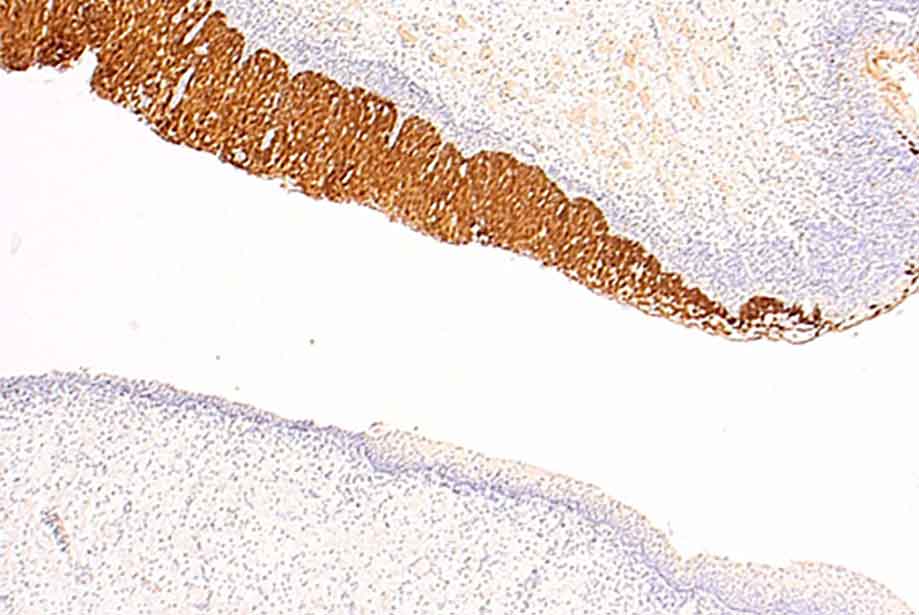
Below is an image of Enzo’s
HIGHDEF® Red IHC Chromogen (AP) used for AP-mediated IHC detection. The system produces a dark red color upon detecting the target antigen of interest. Target antigen being screened below was for CK 5/6 in the squamous cell carcinoma tissue.
Cytokeratins are supportive structural elements in the cell that extend from the nucleus to desmosomes. The overexpression of cytokeratins are attractive target antigens because increased expression is associated with increased mitotic activity.
 for AP-mediated detection systems.")
Figure 2. Formalin-fixed paraffin embedded squamous cell carcinoma human tissue stained with a CK 5/6 antibody labeled with HIGHDEF® Red IHC Chromogen (ADI-950-141) for AP-mediated detection systems.
|
| Chromogens |
Precipitate Color |
Solubility in ethanol/xylene |
Recommended Nuclear Counterstain |
Characteristics |
| BCIP/NBT |
Dark Blue |
insoluble |
Neutral Red |
- coarse granular stain
- longer incubation times than diazonium-based substrates/higher stability
- do not experience turbidity like diazonium salts, less background
- no significant differences in sensitivity with other substrates
- intense color
|
| BCIP/T-NBT |
Brownish to Purple |
insoluble |
Hematoxylin |
- fine granular stain
- longer incubation times than diazonium-based substrates/higher stability
- do not experience turbidity like diazonium salts, less background
- no significant differences in sensitivity with other substrates
- intense color
|
| Naphthol ASMXP + Fast Red |
Red/Orange |
soluble |
Hematoxylin |
- amorphous
- no significant differences in sensitivity with other substrates
- experience slight turbidity with longer incubations
- prone to fading
- less intense but useful for double staining
|
| Naphthol ASMXP + Fast Blue |
Blue |
soluble |
Neutral Red |
- amorphous
- no significant differences in sensitivity with other substrates
- experience slight turbidity with longer incubations
- prone to fading
- less intense but useful for double staining
|
Table 2. Comparing Common Chromogens used in AP-based IHC systems.
Enzo offers a complete range of products for
Immunohistochemistry detection ranging from
antigen retrieval, to the
mounting medium to prepare your slides. Please check out our
complete range of IHC products here. For more suggestions on immunostaining, please see our
IHC E-book,
10 Tips for Successful IHC ,
Immunohistochemistry Troubleshooting Guide and our
IHC-related application notes. For any further questions and concerns regarding any of our products, please reach out to our Technical Support team. We are here to assist you with your
IHC solutions!

 Kit")