Products related to Coronavirus Biology
SARS-CoV Viral Entry
Monitoring of T Cell Exhaustion
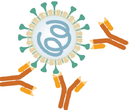
T cells play a critical role in antiviral immunity and their exhaustion has been recently correlated with the progression of the COVID-19 disease. However, it still remains unclear how the viral infection affects the exhaustion of T cells and if it impairs their functional state. We provide a variety of markers to quantify T cells and assess their functional state.
Monitoring of Virion Endocytosis/Lysis
The Endo-/Lysosomal pathway is a key player in viral entry into host cells. Specifically, lysosomal clearance of virions is suspected to be one of the major intracellular defense mechanism against coronaviruses.

See the
Full Infographic
Monitoring of Clathrin-mediated Viral Entry
LYSO-ID® Red cytotoxicity kit (GFP-CERTIFIED®)
Our LYSO-ID Red Cytotoxicity Kit (GFP-CERTIFIED) is the ideal tool to study lysosomal processes and viral biology in fluorescent live microscopy. It monitors dysfunction of lysosomal degradation using a drug-like cationic amphiphilic tracer (CAT) dye that rapidly and selectively stains acidic organelles, and is suitable for monitoring accumulation of lysosomes and lysosome-like structures in live cells.
- Assay includes unique drug-like dye that rapidly partitions into cells and labels acidic organelles
- Only commercial assay available that allows for long-term cell monitoring of cytotoxic effects
- Multi-well, high-throughput with rapid 10-15 minute dye incubation
- No co-incubation with artificial phospholipid analogs required for detection, eliminating the potential for confounding dyeassociated artifacts
- Monitors lysosome accumulation as a response to prolonged drug treatment
- Quantitative results in as little as 3 hours, including drug treatment
Monitoring of Autophagy in SARS-CoV-2 Infection
Autophagy is a self-degradative process that cells use to recycle damaged proteins and destroy pathogens, including SARS-CoV-2. The scientific communities are trying to shed a light on the interplay between the autophagy machinery and SARS-CoV-2 infection but until now it is still not clear if autophagy inhibition or activation would be more effective against SARS-CoV-2.
 CYTO-ID® Autophagy detection kit 2.0
Autophagy is a self-degradative process that cells use to recycle damaged proteins and destroy pathogens, including SARS-CoV-2. CYTO-ID® Autophagy Detection Kit 2.0 measures autophagic vacuoles and monitors autophagic flux in lysosomally inhibited live cells using a novel dye that selectively labels accumulated autophagic vacuoles. The dye has been optimized through the identification of titratable functional moieties that allow for minimal staining of lysosomes while exhibiting bright fluorescence upon incorporation into pre-autophagosomes, autophagosomes, and autolysosomes (autophagolysosomes). The assay offers a rapid and quantitative approach to monitoring autophagy in live cells without the need for cell transfection.
- No transfection assay eliminates need for transfection efficiency validation
- Brighter, more photostable dye specifically stains autophagic vesicles
- Negligible staining of lysosomes reduces background seen with other dyes
- Rapidly quantifies autophagy in native heterogeneous cell populations
- Facilitates high-throughput screening of activators and inhibitors of autophagy

HeLa cells were stained with CYTO-ID® Green Detection Reagent 2 after being cultured in (A) full media or (B) starvation media (EBSS) with 40μM Chloroquine for 4h. Cells starved in EBSS in the presence of Chloroquine showed very bright green fluorescent signals and punctate structures.
Viral Cytotoxicity Assays
Culture medium - Serum-free medium for the culture of airway cells