Applications of Fluorescence Microscopy
Fluorescence microscopy is a widely practiced technique intended to image characteristic cell population changes over time along with their various cellular and genetic structures. The fluorescent signal used to generate images comes from fluorophore tags bound or associated with target structures such as the
plasma membrane,
mitochondria, and
nuclei. These fluorophores as designed not to interfere with native biological function as much as possible. Standard fluorescence cycles start with the absorption of high energy photons by the fluorophore. This excites the lower energy singlet ground state into another singlet state that is higher in energy which usually lasts on the scale of a few nanoseconds or longer. This excited state eventually returns to the ground state and emits a longer wavelength photon that will be many times lower in brightness compared to the initial excitation. After reaching through a spectral emission filter and detector, the signal is processed for image visualization. By harnessing these principles, researchers can observe mechanistic studies of chemical and biological reactions across cell cultures for extended periods of time.
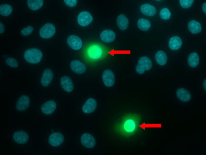
Figure 1: The NUCLEAR-ID® Blue/Green dye (ENZ-53004) is detected as blue-stained nuclei in live cells and fluorescent-green nuclei in dead cells (arrows).
|
Limitations Due to the Photobleaching Effect
However, all fluorescent microscopy techniques will eventually be hindered due to the photobleaching effect. Photobleaching can be defined as the gradual decrease in emitted fluorescence brought by destruction of fluorophores through continual photon excitation. This photochemical alteration is usually the result of covalent bonds breaking and rearranging within the same molecule or with neighboring molecules. Ultimately this destroys the molecule’s fluorogenic properties and results in a continual decrease in image clarity over time. This places a narrow limit on the available window of time researchers can clearly view their stained components.
At this time, much of the intricate details explaining photobleaching are not well understood. This is partially due to each fluorophore having different stabilities and reacting differently in various media. One proposed explanation can be attributed to triple state degradation. While the previously mentioned fluorescence cycle usually involves a singlet state transition, it is also possible to achieve a triplet state excitation. Comparatively, the triplet state is a rarer circumstance. However, even the most robust fluorogenic molecules can only withstand a finite number of absorption-emission cycles before this occurs given enough light exposure. While singlet state transitions can occur in mere nanoseconds, triplet state transitions can last up to microseconds before dropping to the ground state. During this delayed drop, additional photon absorption can increase chemical reactivity and promote covalent bond alterations that lead up to fluorophore destruction.
Fluorescent particle diffusion rate is another factor that may accelerate the photobleaching process faster in some samples compared to others. Faster fluorophore diffusion rates permits shorter time spent under focused light and prevents overall sample fluorescence to degrade as fast. Slower diffusion rates on the other hand forces fluorescent molecules to be closer in vicinity and can accelerate this destruction. Slower diffusion rates can result from larger molecular complexes impeding migration, such as protein complexes or vesicles as examples. More viscous media such as gels and concentrated solutions also play a role in limiting movement. Some fluorophores are inherently more prone to photobleaching than others due to their photon absorption properties. For example, red colored fluorophores tend to have excitation wavelength ranges that overlap with green variants. In multi-color live cell imaging, this problem is especially pronounced since the wider excitation spectra causes’ unnecessary excitation-emission cycles and lead to more rapid degradation.

Figure 2: Fluorescence excitation (Ex 450nm, 570nm) and emission (Em 583nm) spectra for the CYTO-ID® Red Tracer Dye (ENZ-51037).
|
Methods to Overcome Photobleaching
While photobleaching is unavoidable, many have employed practices that can mitigate the rate of degradation. One of the most common optimizations is simply reducing light intensity exposure to fluorophores. Reducing intensity also reduces the frequency of excitation-emission cycles and extends the life of fluorescent molecules. However, reducing light intensity can also reduce emission signal. A careful balance must be found so that image quality is not compromised. Reducing time exposure to light sources also reduces photobleaching as this also decreases the frequency of excitation-emission cycles. Fluorogenic molecules must be stored away from light sources as much as possible until they are ready for viewing. In multi-color studies, selecting dyes with greater photo stability and fewer spectral overlaps can also extend the time window for observation.
Another strategy is to reduce or deplete oxygen in samples. Since triple state transitions are known to break and rearrange covalent bonds, reacting with molecular oxygen can create reactive oxygen species. This is not only a hazard to cellular health, but also damages fluorescent molecules. Depleting or reducing O2 mitigates this possibility and encourages longer lasting fluorescence. One popular oxygen scavenging system employs glucose oxidase and catalase (GOC). Reactive oxygen species can also be reduced by incorporating a number of antioxidants such as ascorbic acid or n-Propyl gallate (nPG) as examples. Many
antifade mounting reagents prolong photo stability since they contain reactive oxygen species scavengers and oxygen scavengers. But while oxygen depletion works best for anaerobic organisms, it is much less effective in mammalian cells or tissue samples due to negative impacts towards normal cell physiology and health. Adjusting to more frequent pulses of lower-energy photons compared to high-energy photon pulses at lower frequency has also shown significant increases in photo stability. These practices allow image quality to be sustained while also dramatically reducing photobleaching. Delays in excitation provide more time for triplet state transitions to return to the ground state and avoids further decay.
At Enzo Life Sciences, we’ve translated our expertise in fluorescent probe chemistry and cellular analysis into our
CELLESTIAL® portfolio. From simple organelle-specific dyes for imaging
cell structure and determining
cell viability, to more complex dyes and reporter assays for monitoring cell signal,
death pathways, and
toxicity, every product is developed and reliably manufactured to provide sensitivity, specificity, and convenience. We also offer
gold standard dyes and
EQ quencher dyes for FRET applications. Enzo’s
Spectra Viewer provides clarity on corresponding excitation and emission for a variety of live-cell analysis kits and dyes. Check out our
Successful Research Tips or contact our
Technical Support Service for further assistance.


