Enzo Life Sciences provides over 40 years of experience in the manufacturing and supply of research kits, biochemicals and biologicals. As
Scientists Enabling Healthcare™, we realize the value in providing relevant information to our customers working in the fields of life sciences, drug development and clinical research. We are happy to share simple but useful hints for improving your daily tasks as well as the overall quality of your results. With this in mind, below is a list of tips for achieving high quality data by fluorescent
in situ hybridization (FISH). This list of suggestions is based not only on Enzo’s experience as a recognized pioneer in labeling and detection technologies, but also on solutions we offer on a regular basis, in order to assist researchers in obtaining the most accurate and consistent results.
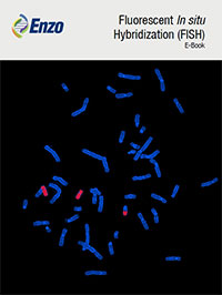
FISH (Fluorescent In Situ hybridization)
Fluorescent
in situ hybridization (FISH) is a cytogenetic technique enabling "mapping" of the genetic material of human cells. It is commonly used to label DNA providing information on the location, length, and number of copies of specific genes or chromosome portions. Additionally, it can be applied to all types of RNA, and is foremost utilized to detect copy numbers and location of mRNA to visualize cellular transcription activity. FISH is based on the specific interaction between a fluorescence-labeled probe and a specific target sequence in cellular DNA or RNA.
Select the right bait for FISH-ing
Ready-to-use probes are commercially available, but they can be quite expensive and do not always offer the required experimental flexibility. One option is to synthetize your own probes. In general, double-stranded DNA probes are easy to prepare, label, and work with in the laboratory, while single-stranded RNA probes are uniform in size, achieve high incorporation of label, and form highly stable RNA-RNA hybrids. DNA probes are usually prepared by nick translation (from supercoiled or linear DNA) or random priming (from linearized DNA); riboprobes can be obtained by in vitro transcription from linearized vectors containing RNA polymerase promotors. Taking all this into account, you can decide what kind of probe is best suited for your needs (DNA- or RNA-based), depending on your starting material (type, quantity, quality) and the tools available.

Quality of the input DNA
The quality of the probe is vital for successful FISH and this is in turn strictly related to the quality of the template DNA. Make sure to use good quality, purified DNA, free of RNA, protein, or other contaminants; for example, use a DNA purification procedure that selectively purifies plasmid DNA (e.g., anion exchange column-based purification). The purity of the preparation can be determined using classical gel electrophoresis, automated electrophoresis systems, or a spectrophotometric system (such as a NanoDrop).
Quality of the probe
Depending on the application, you may need to purify the probe in order to remove unincorporated nucleotides. The yield, dye incorporation and fragment length should be verified. All these elements will give you a good insight on the quality of the probes. For example, if RNA probes appear as a sharp band on an agarose gel, DNA probes will most likely form a smear, generally with the majority of fragments between 100 and 250 bp. If obtaining low yield, inefficient dye incorporation or unexpected probes length, you might need to optimize the previous steps: quality of the template, amount of the starting material, reaction temperatures and time, etc.
Sample preparation
The quality of test specimens is critical for obtaining reliable and consistent results. Tissues can be fixed with formalin or paraformaldehyde, whereas precipitating fixatives (e.g. alcohols) should be avoided to correctly preserve nucleic acids. In general, do not exceed 24 hours of fixation (at maximum), as this would make probe penetration more difficult and increase auto-florescence. Sections should be 3-4μm thick – thicker slices can lead to problems in probe penetration, as well as in the interpretation of the results because of different focal planes; too thin sections can instead truncate the signal and make the manipulation more difficult. For sample preparations, all tools should be treated with alcohol and/or DNAse/RNAse eliminating agents, especially if unfixed or fixed, since cryopreserved samples are processed with the same equipment.
Cells for DNA FISH, on the other hand, should be treated with a hypotonic solution (e.g. sodium citrate and BSA) and then fixed, usually with a 3:1 methanol/acetic acid solution (freshly prepared). Try to distribute cells uniformly across the slide, to avoid clumps and nuclei overlapping. Cell density can be adjusted by centrifuging and resuspending them in a higher or lower volume. Once on the slide, the cytoplasm should not be visible, as this could interfere with the hybridization. If that is the case, pepsin (or other peptidase treatments) might be used; alternatively, try to re-fix the cells in fresh solution.
Prepare the slide
Pre-clean the glass slides with 70% ethanol before use. If needed, treat them in order to render their surface adhesive (e.g. with poly-lysine).
Pre-treatment of specimens
Choose the appropriate pre-treatment in order to allow the subsequent hybridization. On the basis of sample type, the tools, and the time at your disposal, evaluate the most suitable protocol for proper dewaxing (for FFPE sections), sample permeabilization (using proteases, detergents, alcohols, etc), denaturation of the probes and target (notably for dsDNA probes, using pH or heat), slides aging in case of cell spreads, etc.

Hybridization
Hybridization specificity (stringency) is driven by the degree of complementarity between the probe and target sequences, as well as by the probe length. These characteristics will directly influence probe concentration, temperature and time of hybridization, and concentration of monovalent cations present in the hybridization solution needed to obtain the best results. Careful tuning of these parameters can help to remove non-specific interactions. Hybridization temperature, typically ranging between 55 and 62 °C, is probably the most important variable to consider when aiming for high specificity. Among the components of the hybridization buffer, formamide allows hybridization at temperatures significantly lower than the actual melting temperature of a probe-target-hybrid and thus may assist in the conservation of the morphology of samples. In addition, Cot DNA sequences are routinely included to reduce non-specific hybridization to repetitive DNA sequences. In this phase, it is also important to keep the humidity conditions under control, in order to avoid drying out or overconcentration of the solution. This could interfere with the hybridization, lead to high fluorescent background or to altered chromosome morphology.
Post hybridization
The post hybridization washes are as important as the hybridization itself to assure specificity. Gradually increasing the stringency will remove weaker (and likely less specific) interactions between the probe and the target. To set up optimal conditions, take the type of probe you are using into account: RNA-DNA hybrids are more stable than DNA-DNA hybrids.
Mount and visualize
Use preferentially an antifade mounting medium and DAPI as a counterstaining. Do not expose the sample to high light intensity for too long, in order to reduce photobleaching. When enumerating signals in metaphase nuclei or chromosomes spreads, just take intact, clearly separated chromosomes/nuclei, with non-overlapping signals into account. Carefully evaluate doubtful cases at higher magnification. In general, split signals (e.g. small fluorescent spots close to each other) can be counted as one. Repeat counting for more accuracy in case of uncertainties.

Avoid contamination
Change the solutions frequently and use preferably dedicated jars for FISH reagents; wash the material frequently. Pay attention when pipetting the probe to avoid the pipette touching the tube and remember to change pipette tips at each step. It can be beneficial to treat materials as well as the work area with DNAse/RNAse eliminating agents.
Enzo Life Sciences is a global leader in DNA and RNA labeling technologies. We offer a range of products for your
Genomics and
Cancer research needs. For a simple and efficient method for generating labeled DNA, please check out our
Nick translation DNA labeling kit as well as a list of our
SEEBRIGHT® fluorescent dye-dUTPs. Don’t forget to check Enzo’s
spectra viewer for excitation and emission wavelength profiles of common dyes and our other fluorescence cell-based assays. While you’re at it, please check out our TechNote on the
pros and cons of FISH, aCGH, and NGS. For all questions and concerns regarding any of our products, our
Technical Support Team is here to assist.