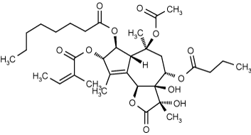
Replaces Prod. #: ALX-350-004
- Potent inhibitor of SERCA
- Cell-permeable tumor promoter
Thapsigargin is a potent inhibitor of sarco/endoplasmic reticulum Ca2+-ATPases (SERCA), which are linked with endoplasmic reticulum stress, and induces the release of intracellular stored Ca2+ without hydrolysis of inositolphospholipid (IC50=30nM). Inhibition of SERCA by thapsigargin reveals a significant change in intracellular Ca2+ homeostasis and pH regulation in tumor cells, and may be used to distinguish between discrete intracellular Ca2+ pools. Thapsigargin increases Ca2+-dependent Na+ influx in human platelets in a dose-dependent manner. Thapsigargin-induced tumor promotion and down-regulation of the EGF receptor is independent of PKC activation. Thapsigargin also stimulates nitric oxide (NO) production and as a result, may promote apoptosis.
Product Details
| Formula: | C34H50O12 |
| |
| MW: | 650.8 |
| |
| Source: | Isolated from Thapsia garganica. |
| |
| CAS: | 67526-95-8 |
| |
| MI: | 14: 9272 |
| |
| Purity: | ≥95% (HPLC, TLC) |
| |
| Appearance: | Colorless wax or white solid. |
| |
| Solubility: | Soluble in acetone, DMSO (20mg/ml), or 100% ethanol (20mg/ml). |
| |
| Shipping: | Ambient Temperature |
| |
| Long Term Storage: | -20°C |
| |
| Use/Stability: | Stable for at least 1 year after receipt when stored, as supplied, at -20°C. |
| |
| Handling: | USE CAUTION – product is a tumor promoter. Solutions may lose activity after one week. For longer term storage, dissolve in acetone or ethanol, aliquot and evaporate solvent to dryness with a stream of nitrogen or argon. Store aliquots of neat compound at -20°C or colder. Protect from light and oxygen. |
| |
| Technical Info/Product Notes: | Replacement for ADI-908-298. |
| |
| Regulatory Status: | RUO - Research Use Only |
| |
Please mouse over
Product Literature References
PERK recruits E-Syt1 at ER–mitochondria contacts for mitochondrial lipid transport and respiration: M.L. Sassano, et al.; J. Cell Biol.
222, e202206008 (2023),
Abstract;
A non-invasive system to monitor in vivo neural graft activity after spinal cord injury: K. Ago, et al.; Commun. Biol.
5, 803 (2022),
Abstract;
Ca2+-mediated mitochondrial inner membrane permeabilization induces cell death independently of Bax and Bak: G. Quarato, et al.; Cell Death Differ.
29, 1318 (2022),
Abstract;
Diverse maturity-dependent and complementary anti-apoptotic brakes safeguard human iPSC-derived neurons from cell death: R. Wilkens, et al.; Cell Death Dis.
13, 887 (2022),
Abstract;
Full Text
ER stress-induced cell death proceeds independently of the TRAIL-R2 signaling axis in pancreatic β cells: C. Hagenlocher, et al.; Cell Death Dis.
8, 34 (2022),
Abstract;
The alkalinizing, lysosomotropic agent ML-9 induces a pH-dependent depletion of ER Ca2+ stores in cellulo: M. Kerkhofs, et al.; Biochim. Biophys. Acta Mol. Cell Res.
1869, 119308 (2022),
Abstract;
ACRBP (Sp32) is involved in priming sperm for the acrosome reaction and the binding of sperm to the zona pellucida in a porcine model: Y. Kato, et al.; PLoS One
16, e0251973 (2021),
Abstract;
Full Text
An inhibitor-mediated beta-cell dedifferentiation model reveals distinct roles for FoxO1 in glucagon repression and insulin maturation: T. Casteels, et al.; Mol. Metab.
54, 101329 (2021),
Abstract;
Full Text
ATPase inhibitory factor-1 disrupts mitochondrial Ca2+ handling and promotes pathological cardiac hypertrophy through CaMKIIδ: M. Pavez-Giani, et al.; Int. J. Mol. Sci.
22, 4427 (2021),
Abstract;
Full Text
Blockade of Oncogenic NOTCH1 with the SERCA Inhibitor CAD204520 in T-cell Acute Lymphoblastic Leukemia: M. Marchesini, et al.; Cell Chem. Biol.
27, 678 (2021),
Abstract;
Full Text
Deletion of mFICD AMPylase alters cytokine secretion and affects visual short-term learning in vivo: N. McCaul, et al.; J. Biol. Chem.
297, 100991 (2021),
Abstract;
Selenoprotein DIO2 is a regulator of mitochondrial function, morphology and UPRmt in human cardiomyocytes: N. Bomer, et al.; Int. J. Mol. Sci.
22, 11906 (2021),
Abstract;
Full Text
A molecular mechanism for turning off IRE1α signaling during endoplasmic reticulum stress: X. Li, et al.; Cell Rep.
33, 108563 (2020),
Abstract;
Full Text
Functional peroxisomes are essential for efficient cholesterol sensing and synthesis: K. Charles, et al.; Front Cell Dev Biol
8, 560266 (2020),
Abstract;
Full Text
Proteome instability is a therapeutic vulnerability in mismatch repair deficient cancer: D. McGrail, et al.; Cancer Cell
37, 371 (2020),
Abstract;
Full Text
MICU1 Confers Protection from MCU-Dependent Manganese Toxicity: J. Wettmarchausen, et al.; Cell Rep.
25, 1425 (2018),
Abstract;
Remodeling the endoplasmic reticulum proteostasis network restores proteostasis of pathogenic GABAA receptors: Y.L. Fu, et al.; PLoS One
13, e0207948 (2018),
Abstract;
Spatial Separation of Mitochondrial Calcium Uptake and Extrusion for Energy-Efficient Mitochondrial Calcium Signaling in the Heart: S. De La Fuente, et al.; Cell Rep.
24, 3099 (2018),
Abstract;
Drug-perturbation-based stratification of blood cancer: S. Dietrich, et al.; J. Clin. Invest.
128, 427 (2017),
Abstract;
Full Text
Endocytosis regulates TDP-43 toxicity and turnover: G. Liu, et al.; Nat. Commun.
8, 2092 (2017),
Abstract;
Full Text
Agonist-Mediated Activation of STING Induces Apoptosis in Malignant B Cells: C.A. Tang, et al.; Cancer Res.
76, 2137 (2016),
Application(s): Stimulated cells,
Abstract;
Full Text
AMPK-independent inhibition of human macrophage ER stress response by AICAR: M. Boß, et al.; Sci. Rep.
6, 32111 (2016),
Application(s): Cell culture, assessing ER stress responses in macrophages,
Abstract;
Full Text
Comparative effects of nodularin and microcystin-LR in zebrafish: 2. Uptake and molecular effects in eleuthero-embryos and adult liver with focus on endoplasmic reticulum stress: S. Faltermann, et al.; Aquat. Toxicol.
171, 77 (2016),
Application(s): Positive control for ER-stress induction,
Abstract;
HERPUD1 protects against oxidative stress-induced apoptosis through downregulation of the inositol 1,4,5-trisphosphate receptor: F. Paredes, et al.; Free Radic. Biol. Med.
90, 206 (2016),
Application(s): Cell culture ,
Abstract;
Stromal Interaction Molecule 1 Rescues Store-Operated Calcium Entry and Protects NG115-401L Cells Against Cell Death Induced by Endoplasmic Reticulum and Mitochondrial Oxidative Stress: C. Zhang, et al.; Neurochem. Int.
97, 137 (2016),
Application(s): Cell culture,
Abstract;
Camphene isolated from essential oil of Piper cernuum (Piperaceae) induces intrinsic apoptosis in melanoma cells and displays antitumor activity in vivo: N. Girola, et al.; Biochem. Biophys. Res. Commun.
467, 928 (2015),
Application(s): Cell Culture,
Abstract;
ER localization is critical for DsbA-L to Suppress ER Stress and Adiponectin Down-Regulation in Adipocytes: M. Liu, et al.; J. Biol. Chem.
290, 10143 (2015),
Application(s): Cell Culture,
Abstract;
Full Text
Stress of endoplasmic reticulum modulates differentiation and lipogenesis of human adipocytes: M. Koc, et al.; Biochem. Biophys. Res. Commun.
460, 684 (2015),
Application(s): Cell Culture,
Abstract;
Transient receptor potential canonical 1 (TRPC1) channels as regulators of sphingolipid- and VEGF receptor expression: implications for thyroid cancer cell migration and proliferation: M.Y. Asghar, et al.; J. Biol. Chem.
290, 16116 (2015),
Application(s): Cell Culture,
Abstract;
Full Text
Unfolded protein response signaling by transcription factor XBP-1 regulates ADAM10 and is affected in Alzheimer's disease: S. Reinhardt, et al.; FASEB J.
28, 978 (2014),
Abstract;
Autophagy-mediated insulin receptor down-regulation contributes to endoplasmic reticulum stress-induced insulin resistance: L. Zhou, et al.; Mol. Pharmacol.
76, 596 (2009),
Abstract;
Thapsigargin, a selective inhibitor of sarco-endoplasmic reticulum Ca2+ -ATPases, modulates nitric oxide production and cell death of primary rat hepatocytes in culture: N.K. Canova, et al.; Cell Biol. Toxicol.
23, 337 (2007),
Abstract;
Changes in intracellular Ca2+ and pH in response to thapsigargin in human glioblastoma cells and normal astrocytes: G.G. Kovacs, et al.; Am. J. Physiol. Cell. Physiol.
289, C361 (2005),
Abstract;
Thapsigargin induces a calmodulin/calcineurin-dependent apoptotic cascade responsible for the death of prostatic cancer cells: B. Tombal, et al.; Prostate
43, 303 (2000),
Abstract;
Thapsigargin induces apoptosis in cultured human aortic smooth muscle cells: C. Peiro, et al.; J. Cardiovasc. Pharmacol.
36, 676 (2000),
Abstract;
Nitric oxide is involved in apoptosis induced by thapsigargin in rat mesangial cells: A.M. Rodriguez-Lopez, et al.; Cell Physiol. Biochem.
9, 285 (1999),
Abstract;
Signal transduction of thapsigargin-induced apoptosis in osteoblast: H.J. Chae, et al.; Bone
25, 453 (1999),
Abstract;
Baculovirus p35 and Z-VAD-fmk inhibit thapsigargin-induced apoptosis of breast cancer cells: X.M. Qi, et al.; Oncogene
15, 1207 (1997),
Abstract;
Role of EGR-1 in thapsigargin-inducible apoptosis in the melanoma cell line A375-C6: S. Muthukkumar, et al.; Mol. Cell. Biol.
15, 6262 (1995),
Abstract;
Full Text
Intracellular Ca2+ signals activate apoptosis in thymocytes: studies using the Ca(2+)-ATPase inhibitor thapsigargin: S. Jiang, et al.; Exp. Cell Res.
212, 84 (1994),
Abstract;
The role of calcium, pH, and cell proliferation in the programmed (apoptotic) death of androgen-independent prostatic cancer cells induced by thapsigargin: Y. Furuya, et al.; Cancer Res.
54, 6167 (1994),
Abstract;
Thapsigargin, a Ca(2+)-ATPase inhibitor, depletes the intracellular Ca2+ pool and induces apoptosis in human hepatoma cells: A. Tsukamoto & Y. Kaneko; Cell Biol. Int.
17, 969 (1993),
Abstract;
Brefeldin A, thapsigargin, and AIF4- stimulate the accumulation of GRP78 mRNA in a cycloheximide dependent manner, whilst induction by hypoxia is independent of protein synthesis: B.D. Price, et al.; J. Cell. Physiol.
152, 545 (1992),
Abstract;
Demonstration of two forms of calcium pumps by thapsigargin inhibition and radioimmunoblotting in platelet membrane vesicles: B. Papp, et al.; J. Biol. Chem.
266, 14593 (1991),
Abstract;
Identification of intracellular calcium pools. Selective modification by thapsigargin: J.H. Bian, et al.; J. Biol. Chem.
266, 8801 (1991),
Abstract;
Thapsigargin, a tumor promoter, discharges intracellular Ca2+ stores by specific inhibition of the endoplasmic reticulum Ca2(+)-ATPase: O. Thastrup, et al.; Proc. Natl. Acad. Sci. USA
87, 2466 (1990),
Abstract;
Activation of calcium entry by the tumor promoter thapsigargin in parotid acinar cells. Evidence that an intracellular calcium pool and not an inositol phosphate regulates calcium fluxes at the plasma membrane: H. Takemura, et al.; J. Biol. Chem.
264, 12266 (1989),
Abstract;
Phosphorylation at threonine-654 is not required for negative regulation of the epidermal growth factor receptor by non-phorbol tumor promoters: B.A. Friedman, et al.; Proc. Natl. Acad. Sci. USA
86, 812 (1989),
Abstract;
A novel tumour promoter, thapsigargin, transiently increases cytoplasmic free Ca2+ without generation of inositol phosphates in NG115-401L neuronal cells: T.R. Jackson, et al.; Biochem. J.
253, 81 (1988),
Abstract;
Thapsigargin, a novel promoter, phosphorylates the epidermal growth factor receptor at threonine 669: K. Takishima, et al.; Biochem. Biophys. Res. Commun.
157, 740 (1988),
Abstract;
Palytoxin is a non-12-O-tetradecanoylphorbol-13-acetate type tumor promoter in two-stage mouse skin carcinogenesis: H. Fujiki, et al.; Carcinogenesis
7, 707 (1986),
Abstract;
General Literature References
A tool coming of age: thapsigargin as an inhibitor of sarco-endoplasmic reticulum Ca(2+)-ATPases: M. Treiman, et al.; TIPS
19, 131 (1998), (Review),
Abstract;
The sarcoplasmic reticulum Ca2+ pump: inhibition by thapsigargin and enhancement by adenovirus-mediated gene transfer: G. Inesi, et al.; Ann. NY Acad. Sci.
853, 195 (1998), (Review),
Abstract;
Full Text
Use of thapsigargin to study Ca2+ homeostasis in cardiac cells: T.B. Rogers, et al.; Biosci. Rep.
15, 341 (1995), (Review),
Abstract;
Thapsigargin, a high affinity and global inhibitor of intracellular Ca2+ transport ATPases: G. Inesi and Y. Sagara; Arch. Biochem. Biophys.
298, 313 (1992), (Review),
Abstract;
Role of Ca2(+)-ATPases in regulation of cellular Ca2+ signalling, as studied with the selective microsomal Ca2(+)-ATPase inhibitor, thapsigargin: O. Thastrup; Agents Actions
29, 8 (1990), (Review),
Abstract;
Related Products


