Product Details
| Alternative Name: | Proteasome subunit α type-1, -2, -3, -4, -5 & -6, Macropain subunits C2, C3, C8, C9, ι & ζ |
| |
| Clone: | MCP231 |
| |
| Host: | Mouse |
| |
| Isotype: | IgG1κ |
| |
| Immunogen: | Dinitrophenylated proteasomes. |
| |
| UniProt ID: | P25786 (human PSMA1), P25787 (human PSMA2), P25788 (human PSMA3), P25789 (human PSMA4), P28066 (human PSMA5), P60900 (human PSMA6) |
| |
| Source: | Purified from hybridoma tissue culture supernatant. |
| |
| Species reactivity: | Human, Mouse, Rat
Potato, Rabbit, Yeast
|
| |
| Specificity: | Recognizes the α1, 2, 3, 5, 6 & 7 subunits of the 20S proteasome. |
| |
| Applications: | IHC, WB
|
| |
| Purity Detail: | Protein G affinity purified. |
| |
| Formulation: | Liquid. In PBS containing 0.01% sodium azide. |
| |
| Handling: | Store unopened vial at -20°C. Avoid freeze/thaw cycles. |
| |
| Shipping: | Blue Ice |
| |
| Long Term Storage: | -20°C |
| |
| Scientific Background: | The proteasome is widely recognised as the central enzyme of non-lysosomal protein degradation. It is responsible for intracellular protein turnover and it is also critically involved in many regulatory processes and, in higher eukaryotes, in antigen processing. The 26S proteasome is the key enzyme of the ubiquitin/ATP-dependent pathway of protein degradation. The catalytic core of this unusually large (2000kDa, 450Å in length) complex is formed by the 20S proteasome, a barrel shaped structure shown by electron microscopy to comprise of four rings each containing seven subunits. Based on sequence similarity, all fourteen 20S proteasomal subunit sequences may be classified into two groups, α and β, each group having distinct structural and functional roles. The α-subunits comprise the outer rings and the β-subunits the inner rings of the 20S proteasome. Observations of the eukaryotic proteasome and analysis of subunit sequences indicate that each ring contains seven different subunits (α7β7β7α7) with a member of each sub-family represented in each particle. Each subunit is located in a unique position within the α- or β-rings. 120S Proteasomes degrade only unfolded proteins in an energy-independent manner, whereas 26S proteasomes degrade native and ubiquitinylated proteins in an ATP-dependent manner. The native protein substrates are recognised by subunits, some with ATP binding sites, of the outer 19S caps of the 26S proteasome. The hybridoma secreting the antibody to subunits HC2, HC3, HC8, HC9, Iota and Zeta was generated by fusion of spenocytes from Balb/c mice which had recieved repeated immunisation with dinitrophenylated proteasomes. |
| |
| Technical Info/Product Notes: | Various systems for the nomenclature of the proteasome subunits have been established. This may be a source of confusion as the system on UniProt differs from "standard" nomenclature as described in the literature. The UniProt ID and Gene Name will help to clearly identify the proteins. |
| |
| Regulatory Status: | RUO - Research Use Only |
| |

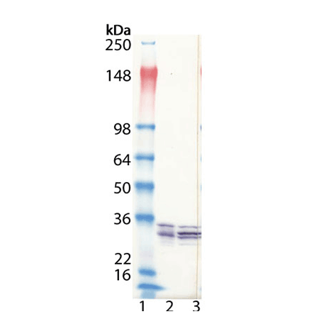
Western Blot analysis of ENZ-ABS667: Lane 1: NovexTMSeeBlueTMPlus2 MW Marker, Lane 2: HeLa cell lysate (Prod. No. ADI-LYC-HL100), Lane 3: Jurkat cell lysate (Prod. No. ADI-LYC-JK100).

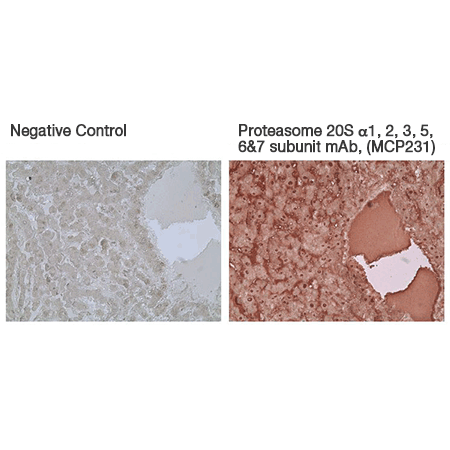
Immunohistochemistry analysis of formalin-fixed, paraffin-embedded human liver tissue stained with Proteasome 20S α1, 2, 3, 5, 6 & 7 subunits monoclonal antibody (MCP231), (Prod. No. ENZ-ABS667) at 10µg/ml.
Please mouse over
General Literature References
Subunit arrangement in the human 20S proteasome: F. Kopp et al.; PNAS
94, 2939 (1997),
Abstract;
The 26S proteasome: subunits and functions: K. Tanaka et al.; Mol. Biol. Rep.
24, 3 (1997),
Abstract;
Human proteasomes analysed with monoclonal antibodies: K. B. Hendil et al.; Biochem. J.
305, 245 (1995),
Abstract;
Related Products


