-
Novel endoplasmic reticulum-selective dye stains live, permeabilized or fixed cells
-
Easily multiplexed with common fluorescent dyes and fluorescent proteins
-
Highly resistant to photo-bleaching, concentration quenching and photo-conversion
-
Stringently manufactured, to control and eliminate non-specific assay artifacts
ER-ID® Green assay kit contains an endoplasmic reticulum-selective dye suitable for live cell, or detergent-permeabilized aldehyde-fixed cell staining. Micromolar concentrations of ER-ID® Green dye are sufficient for staining mammalian cells, as validated with human cervical carcinoma cell line, human T-lymphocyte cell line, Jurkat, HeLa and human bone osteosarcoma epithelial cell line, U2OS. One important application of ER-ID® Green dye is in fluorescence co-localization imaging with red fluorescent protein (RFP)- or orange fluorescent protein (OFP)-tagged proteins, a powerful approach for determining the targeting of molecules to intracellular compartments and for screening of their associations and interactions. However, to date, photoconversion of fluorescent dyes to other colors and metachromatic artifacts, wherein fluorescent dyes emit both in the red (or orange) and green regions of the spectrum, have led to spurious results in co-localization experiments. Additionally, many organelle-targeting probes photobleach rapidly, are subject to quenching upon concentration in organelles, are highly toxic, or only transiently associate with the target organelle, requiring imaging within a minute or two of dye addition. Consequently, ER-ID® Green dye, a new green-emitting, cell-permeable small molecule organic probe that spontaneously localizes to live or fixed endoplasmic reticula, was developed. ER-ID® Green dye can be readily used in combination with other common UV and visible light excitable organic fluorescent dyes and various fluorescent proteins in multi-color imaging and detection applications. The dye emits in the FITC region of the visible light spectrum, and is resistant to photo-bleaching, concentration quenching and photoconversion.The ER-ID® Green assay kit is specifically designed for use with RFP- or OFP-expressing cell lines, as well as cells expressing blue or cyan fluorescent proteins (BFPs, CFPs). Additionally, the kit is suitable for use with live or post-fixed cells in conjunction with probes, such as labeled antibodies, or other fluorescent conjugates displaying similar spectral properties as Texas Red, or coumarin. A nuclear counterstain is provided to highlight this organelle as well.
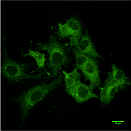
HepG2 cells were fixed with 4% paraformaldehyde for 20 minutes and labeled with ER-ID® for 30 minutes. Image was taken 3 days after the labeling.
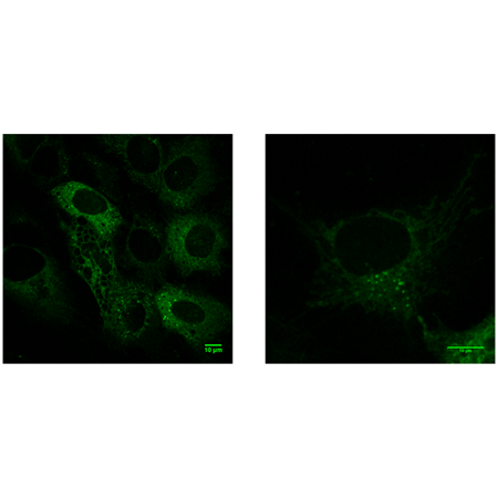
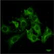
HepG2 cells were incubated 30 min with ER-ID®, according to the protocol, and analyzed with an inverted microscope (Zeiss).
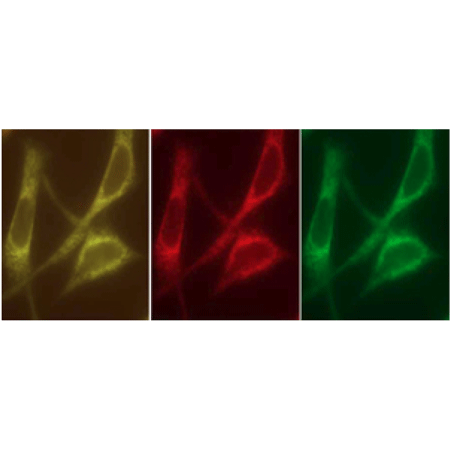
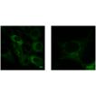
Figure 1: ER-ID® Green staining co-localizes with calreticulin protein containing the KDEL sequence, fused to orange fluorescent protein (OFP).
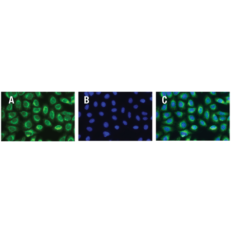
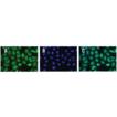
Live HeLa cells stained with ER-ID® Green dye (A), Hoechst 33342 nuclear stain (B) and resulting composite image (C).
Please mouse over
Product Details
| Applications: | Fluorescence microscopy
|
| |
| Application Notes: | Enzo Life Sciences’ ER-ID® Green assay kit contains a novel endoplasmic reticulum-selective dye suitable for live cell, or detergent-permeabilized aldehyde-fixed cell staining. |
| |
| Quality Control: | A sample from each lot of ER-ID® Green assay kit is used to stain HeLa cells, using the procedures described in the user manual. The selectivity of the ER-ID® Green dye is evident. |
| |
| Quantity: | 500 assays |
| |
| Use/Stability: | With proper storage, the kit components are stable up to the date noted on the product label. Store kit at -20˚C in a non-frost free freezer, or –80˚C for longer term storage. |
| |
| Handling: | Protect from light. Avoid freeze/thaw cycles. |
| |
| Shipping: | Blue Ice |
| |
| Long Term Storage: | -20°C |
| |
| Contents: | ER-ID® Green detection reagent: 50μl
Hoechst 33342 nuclear stain: 50μl
10X assay buffer: 15ml |
| |
| Technical Info/Product Notes: | The ER-ID® Green assay kit is a member of the CELLESTIAL® product line, reagents and assay kits comprising fluorescent molecular probes that have been extensively benchmarked for live cell analysis applications. CELLESTIAL® reagents |
| |
| Regulatory Status: | RUO - Research Use Only |
| |
Product Literature References
Molecular interplay between NOX1 and autophagy in cadmium-induced prostate carcinogenesis: A. Tyagi, et al.; Free Radic. Biol. Med.
199, 44 (2023),
Abstract;
Ammonia induces amyloidogenesis in astrocytes by promoting amyloid precursor protein translocation into the endoplasmic reticulum: A. Komatsu, et al.; J. Biol. Chem.
298, 101933 (2022),
Abstract;
PLP1 mutations in patients with multiple sclerosis: identification of a new mutation and potential pathogenicity of the mutations: N.C. Cloake, et al.; J. Clin. Med.
7, 342 (2018),
Application(s): Fluorescence microscopy,
Abstract;
Full Text
Englerin A induces an acute inflammatory response and reveals lipid metabolism and ER stress as targetable vulnerabilities in renal cell carcinoma: A. Batova, et al.; PLoS One
12, e0172632 (2017),
Application(s): Fluorescence microscopy,
Abstract;
Full Text
Cell growth on (“Janus”) density gradients of bifunctional zeolite L crystals: N.S. Kehr, et al.; ACS Appl. Mater. Interfaces
8, 35081 (2016),
Abstract;
The parathyroid hormone second receptor PTH2R and its ligand tuberoinfundibular peptide of 39 residues TIP39 regulate intracellular calcium and influence keratinocyte differentiation: E. Sato, et al.; J. Invest. Dermatol.
136, 1449 (2016),
Abstract;
Viral genome imaging of hepatitis C virus to probe heterogeneous viral infection and responses to antiviral therapies: V. Ramanan, et al.; Virology
494, 236 (2016),
Application(s): Endoplasmic reticulum staining,
Abstract;
Localization-Dependent Cell-Killing Effects of Protoporphyrin (PPIX)-Lipid Micelles and Liposomes in Photodynamic Therapy: S. Tachikawa, et al.; Bioorg. Med. Chem.
23, 7578 (2015),
Application(s): Cell staining,
Abstract;
Related Products


