Product Details
| Alternative Name: | HIAP2, BIRC-2, Baculoviral IAP repeat-containing protein-2, Cellular inhibitor of apoptosis-1, Human inhibitor of apoptosis protein-2, Inhibitor of apoptosis protein-2 |
| |
| Clone: | 1E1-1-10 |
| |
| Host: | Rat |
| |
| Isotype: | IgG2a |
| |
| Immunogen: | Synthetic peptide corresponding to aa 221-232 of mouse c-IAP1. |
| |
| UniProt ID: | Q62210 |
| |
| Species reactivity: | Human, Mouse
|
| |
| Applications: | WB
|
| |
| Recommended Dilutions/Conditions: | Western Blot (1:1,000-1:4,000 using ECL; suggested blocking and dilution buffer is PBST containing 0.1% Tween 20 and either 5% skim milk or 5% BSA. Suggested incubation time is 1 hour at room temperature or overnight at 4°C.)
Suggested dilutions/conditions may not be available for all applications.
Optimal conditions must be determined individually for each application. |
| |
| Application Notes: | Detects a band of ~62kDa by Western blot.
Not recommended for Immunohistochemistry or Immunoprecipitation. |
| |
| Positive Control: | Included. (ALX-803-335-POS. Wild type MEF lysate in SDS loading buffer). Negative control also included (ALX-803-335-NEG. c-Iap1-/- MEF lysate in SDS loading buffer.) |
| |
| Formulation: | Liquid. 0.2μm-filtered solution in PBS containing 0.05% sodium azide. |
| |
| Use/Stability: | Stable for at least 12 months after receipt when stored at -80°C. |
| |
| Handling: | Avoid freeze/thaw cycles. After opening, prepare aliquots and store at -80°C. |
| |
| Shipping: | Dry Ice |
| |
| Short Term Storage: | +4°C |
| |
| Long Term Storage: | -80°C |
| |
| Scientific Background: | All members of the inhibitor of apoptosis proteins (IAP) family contain at least one, but usually three copies of baculovirus IAP repeat (BIR), a ~70-aa zinc binding domain, and upon overexpression suppress apoptosis. The BIR motif is capable of interacting with caspases and occluding their catalytic pockets. Certain IAPs also possess C-terminal RING domains. RING-containing proteins often act as E3 ubiquitin ligases and can catalyse the degradation of both, themselves and selected target proteins through ubiquitylation. So far, eight human IAPs have been identified: Apollon (Bruce), c-IAP1 (HIAP2; MIHB), c-IAP2 (HIAP1; MIHC), ILP-2 (Ts-IAP), Livin (ML-IAP; KIAP), NAIP, Survivin (TIAP) and XIAP (ILP-1; MIHA). c-IAP1 has been shown to inhibit specific caspases. |
| |
| Regulatory Status: | RUO - Research Use Only |
| |

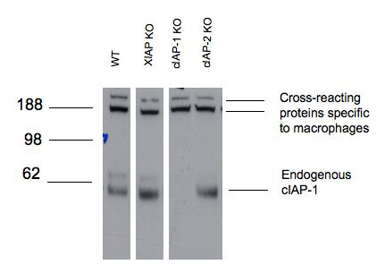
Figure 1: Western blot analysis of c-IAP1 in mouse bone marrow-derived macrophages of the indicated strains lysed in DISC buffer (150mM sodium chloride, 2mM EDTA, 1% Triton X-100, 10% glycerol, 20mM Tris pH 7.5) using MAb to c-IAP1 (1E1-1-10) (Prod. No. ALX-803-335) followed by HRP-anti rat antibody and developed by ECL.Molecular weight marker (kD) is shown at the left. Lanes have been digitally excised from a single blot to aid comparison.

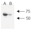
Figure 2: Western blot analysis of human c-IAP1 using MAb to c-IAP1 (1E1-1-10)A: MDA-MB-231 cell lysateB: MDA-MB-231 +LBW 1μM/1h cell lysate (LBW causes degradation of c-IAP1; A. Gaither, et al., 2007)(Image courtesy of Dr. Pascal Meier, The Institute of Cancer Research, London.)

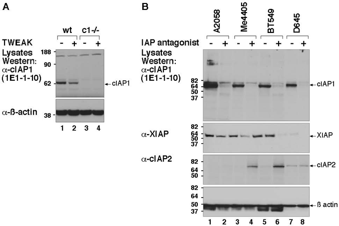
Figure 3: MAb to c-IAP1 (1E1-1-10) is specific for mouse and human c-IAP1. A) Wild type and c-IAP1-/- MEFs were treated +/- TWEAK for 6 hours and whole cell lysates prepared and separated on an SDS-PAGE gel, transferred to a PVDF membrane and probed with MAb to c-IAP1 (1E1-1-10). TWEAK causes a partial loss of c-IAP1. B) 4 human cell lines were treated overnight with an IAP antagonist that induces rapid degradation of c-IAP1. Cells were lysed with DISC lysis buffer and separated on an SDS-PAGE gel, transferred and probed with the MAb to c-IAP1 (1E1-1-10) as in A. XIAP and c-IAP2 blots demonstrate the specificity of the c-IAP1 antibody.
Please mouse over
Product Literature References
XIAP promotes melanoma growth by inducing tumour neutrophil infiltration: M. Daoud, et al.; EMBO Rep.
23, e53608 (2022),
Abstract;
Regulation of CYLD activity and specificity by phosphorylation and ubiquitin-binding CAP-Gly domains: P.R. Elliott, et al.; Cell Rep.
37, 109777 (2021),
Abstract;
Ubiquitin Ligases cIAP1 and cIAP2 Limit Cell Death to Prevent Inflammation: J. Zhang, et al.; Cell Rep.
27, 2679 (2019),
Abstract;
BAX/BAK-Induced Apoptosis Results in Caspase-8-Dependent IL-1β Maturation in Macrophages: D. Chauhan, et al.; Cell Rep.
25, 2354 (2018),
Abstract;
Disruption of XIAP-RIP2 Association Blocks NOD2-Mediated Inflammatory Signaling: T. Goncharov, et al.; Mol. Cell
69, 551 (2018),
Abstract;
Heat Shock Protein 70 (Hsp70) Suppresses RIP1-Dependent Apoptotic and Necroptotic Cascades: S.R. Srinivasan, et al.; Mol. Cancer Res.
16, 58 (2018),
Abstract;
Full Text
Ubiquitin-Mediated Regulation of RIPK1 Kinase Activity Independent of IKK and MK2: A. Annibaldi, et al.; Mol. Cell
69, 566 (2018),
Abstract;
Full Text
X-linked inhibitor of apoptosis protein (XIAP) is a client of heat shock protein 70 (Hsp70) and a biomarker of its inhibition: L.C. Cesa, et al.; J. Biol. Chem.
293, 2370 (2018),
Abstract;
Full Text
Coordinated ubiquitination and phosphorylation of RIP1 regulates necroptotic cell death: M.C. de Almagro, et al.; Cell Death Differ.
24, 26 (2017),
Application(s): Western Blot,
Abstract;
Full Text
Mitochondrial permeabilisation engages NF-κB dependent anti-tumour activity under caspase deficiency: E. Giampazolias, et al.; Nat. Cell Biol.
19, 1116 (2017),
Abstract;
MK2 Phosphorylates RIPK1 to Prevent TNF-Induced Cell Death: I. Jaco, et al.; Mol. Cell
66, 698 (2017),
Abstract;
Full Text
Pentamidine blocks hepatotoxic injury in mice: E. Zhao, et al.; Hepatology
66, 922 (2017),
Abstract;
Full Text
A critical role for cellular inhibitor of protein 2 (cIAP2) in colitis-associated colorectal cancer and intestinal homeostasis mediated by the inflammasome and survival pathways: M. Dagenais, et al.; Mucosal Immunol.
9, 146 (2016),
Abstract;
Activation of TNFR2 sensitizes macrophages for TNFR1-mediated necroptosis: D. Siegmund, et al.; Cell Death Dis.
7, e2375 (2016),
Abstract;
Full Text
Cellular IAP proteins and LUBAC differentially regulate necrosome-associated RIP1 ubiquitination: M.C. de Almagro, et al.; Cell Death Dis.
6, e1800 (2015),
Abstract;
Full Text
Effects of physiological and synthetic IAP antagonism on c-IAP-dependent signaling: A.J. Kocab, et al.; Oncogene
34, 5472 (2015),
Abstract;
RIPK3 promotes cell death and NLRP3 inflammasome activation in the absence of MLKL: K.E. Lawlor, et al.; Nat. Commun.
6, 6282 (2015),
Abstract;
Full Text
An inactivating caspase 11 passenger mutation originating from the 129 murine strain in mice targeted for c-IAP1: N.S. Kenneth, et al.; Biochem. J.
443, 355 (2012),
Application(s): WB using mouse embryonic fibroblast (MEF) cell lysate,
Abstract;
Full Text
Molecular determinants of Smac mimetic induced degradation of cIAP1 and cIAP2: M. Darding, et al.; Cell Death Differ.
18, 1376 (2011),
Abstract;
Full Text
TAK1 is required for survival of mouse fibroblasts treated with TRAIL, and does so by NF-kappaB dependent induction of cFLIPL: J.M. Lluis, et al.; PLoS One
5, e8620 (2010),
Abstract;
Full Text
TNF signaling, but not TWEAK triggered cellular Inhibitor of Apoptosis protein 1 (cIAP1) degradation, requires cIAP1 RING dimerization and E2 binding: R. Feltham, et al.; J. Biol. Chem.
285, 17525 (2010),
Abstract;
Full Text
Cellular IAPs inhibit a cryptic CD95-induced cell death by limiting RIP1 kinase recruitment: P. Geserick, et al.; J. Cell Biol.
187, 1037 (2009),
Abstract;
Full Text
TRAF2 must bind to cellular inhibitors of apoptosis for tumor necrosis factor (tnf) to efficiently activate nf-{kappa}b and to prevent tnf-induced apoptosis: J.E. Vince, et al.; J. Biol. Chem.
284, 35906 (2009),
Abstract;
TWEAK-FN14 signaling induces lysosomal degradation of a cIAP1-TRAF2 complex to sensitize tumor cells to TNFalpha: J.E. Vince, et al.; J. Cell. Biol.
182, 171 (2008),
Abstract;
Full Text
A Smac mimetic rescue screen reveals roles for inhibitor of apoptosis proteins in tumor necrosis factor-alpha signaling: A. Gaither, et al.; Cancer Res.
67, 11493 (2007),
Abstract;
Full Text
IAP antagonists target cIAP1 to induce TNFalpha-dependent apoptosis: J.E. Vince, et al.; Cell
131, 682 (2007),
Abstract;
Determination of cell survival by RING-mediated regulation of inhibitor of apoptosis (IAP) protein abundance: J. Silke, et al.; PNAS
102, 16182 (2005),
Abstract;
Full Text
General Literature References
Destabilizing influences in apoptosis: sowing the seeds of IAP destruction: S.J. Martin; Cell
109, 793 (2002), Review,
Abstract;
Related Products


