Cell permeable fluorescent reagent (Ex(max): 490nm; Em(max): 515nm) for the detection of highly reactive oxygen species (hROS). Immediately reacts with hROS such as hydroxyl radical and peroxynitrite, and the fluorescence intensity greatly increases. In addition, peroxynitrite can be detected in distinction from nitric oxide and superoxide since HPF does not react with nitric oxide, superoxide and hydrogen peroxide. Moreover, HPF is resistant to light-induced autooxidation. HPF does not react with hypochlorite (-OCl) either and thus can be used in combination with APF (Prod. No. ALX-620-075) which detects -OCl to elucidate reliably the roles of -OCl in biological systems such as neutrophils. Moreover, HPF is resistant to light-induced autooxidation. Not for sale in Japan.
Product Details
| Alternative Name: | Hydroxyphenyl fluorescein, 2-[6-(4'-Hydroxy)phenoxy-3H-xanthen-3-on-9-yl]benzoic acid |
| |
| Formula: | C26H16O6 |
| |
| MW: | 424.4 |
| |
| CAS: | 359010-69-8 |
| |
| Concentration: | ~5mM |
| |
| Formulation: | Dissolved in 0.47ml dimethylformamide. |
| |
| Purity: | ≥98% (HPLC) |
| |
| Appearance: | Pale yellow. |
| |
| Shipping: | Blue Ice |
| |
| Long Term Storage: | +4°C |
| |
| Use/Stability: | Prepare 500-5'000-fold dilution (~10-1µM) in phosphate buffer (0.1M phosphate, pH 7.4) immediately before use. BSA, phenol red and amines may affect the fluorescence and must be used with caution. Do not store the dilutions. |
| |
| Handling: | After opening, prepare aliquots and store at +4°C. Protect from light. Keep under inert gas. |
| |
| Protocol: | Precautions for use
1. Prepare the diluted solution just before use and use it up.
2. The dilution buffer should be adjusted to pH 7.0-7.5. Please note that bovine serum albumin (BSA) and phenol red might interfere with the fluorescence measurement.
Light-induced autoxidation (Figure 3)
After adding HPF or DCFH-DA (10µM), the cells were incubated for 30 min at 37°C in the dark. The fluorescence images were obtained with a confocal fluorescent microscope (excitation wavelength: 488nm, fluorescence emission wavelength: using a 515nm barrier filter). The cells were laser-irradiated again at 488nm for 10 sec and fluorescence images were obtained again (see Figure 3).
Fluorescence images of HPF- or APF-loaded neutrophils (Figure 4)
Neutrophils were obtained from porcine blood and suspended in Krebs-Ringer phosphate buffer (114mM NaCI, 4.6mM KCI, 2.4mM MgSO4, 1.0mM CaCI2, 15mM NaH2PO4/Na2HPO4, pH 7.4) and kept on ice until use. Then they were seeded onto a glass-bottomed dish. The cells were loaded with HPF or APF (10µM) by incubation for 30min at room temperature and then stimulated with PMA (including 2ng/ml; 0.1% DMF as a cosolvent). Fluorescence images were obtained with a confocal fluorescent microscope before and 10 min after the stimulation with PMA (excitation wavelength: 488nm, fluorescence emission wavelength: 505-550nm; using a 505-550nm barrier filter).
Detection of hROS in the HRP/H2O2 system using HPF and APF (Figure 5)
Reaction timecourse
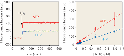
Fluorescence probe reagents (final 10µM; 0.1% DMF as a cosolvent) were added to sodium phosphate buffer (0.1M; pH 7.4) containing HRP (0.2µM). H2O2 (final 1µM) was added at the time indicated by the arrow. The fluorescence intensities were measured at excitation wavelength of 490nm and fluorescence emission wavelength of 515nm. Relation between the amount of added H2O2 and fluorescence increase in the HRP/H2O2 system.
Relation between the amount of added H2O2 and fluorescence increase in the HRP/H2O2 system
Fluorescence probe reagents (final 10µM; 0.1% DMF as a cosolvent) were added to sodium phosphate buffer (0.1M; pH 7.4) containing HRP (0.2µM). The fluorescence intensities were measured at excitation wavelength of 490nm and fluorescence emission wavelength of 515nm. |
| |
| Regulatory Status: | RUO - Research Use Only |
| |

Figure 1: Reaction of Hydroxyphenyl Fluorescein (HPF) with hROS.

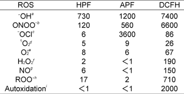
Figure 6: Fluorescence probe reagents were added to sodium phosphate buffer (0.1M, pH 7.4) (final 10µM; 0.1% DMF as a cosolvent). The fluorescence intensities of HPF, APF and DCFH were measured at excitation wavelength of 490, 490 and 500nm and fluorescence emission wavelength of 515, 515 and 520nm, respectively.
a 100µM of Ferrous perchlorate (ll) and 1mM of H2O2 were added.
b 3µM (final) of ONOO- was added.
c 3µM (final) of NaOCI was added.
d 100µM of 3-(1,4-Dihydro-1,4-epidioxy-1-naphthyl)propionic acid was added.
e 100µM of KO2 was added.
f 100µM of H2O2 was added.
g 100µM of 1-Hydroxy-2-oxo-3-(3-aminopropyl)-3-methyl-1-triazene was added.
h 100µM of 2,2

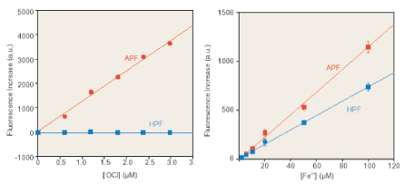
Figure 2: HPF and APF enable the selective detection of highly reactive oxygen species and they are hardly autoxidized by light irradiation. Left panel: detection of -OCl.Right panel: detection of •OH generated by the Fenton reaction.

Figure 3: Light-induced autoxidation. Compared with DCFH, HPF and APF were hardly autoxidized by light irradiation.

Figure 5: Detection of hROS in the HRP/H2O2 system using HPF and APF. The fluorescence intensity increased immediately upon the addition of H2O2. Further, it was found that HPF and APF could detect hROS generated in the HRP/H2O2 system in a dose-dependent manner.

Figure 4: Fluorescence images of HPF- or APF-loaded neutrophils. The fluorescence intensity of HPF-loaded neutrophils was not changed upon stimulation with PMA whereas that of APF-loaded neutrophils greatly increased. This suggests that -OCI produced by MPO (myeloperoxidase) upon stimulation with PMA could be selectively visualized in distinction from other ROS.
Please mouse over
Product Literature References
Gas Flow Shaping via Novel Modular Nozzle System (MoNoS) Augments kINPen-Mediated Toxicity and Immunogenicity in Tumor Organoids: J. Berner, et al.; Cancers
15, 1254 (2023),
Abstract;
Modeling Gas Plasma-Tissue Interactions in 3D Collagen-Based Hydrogel Cancer Cell Cultures: L. Miebach, et al.; Bioengineering (Basel)
10, 367 (2023),
Abstract;
Development of novel fluorescence probes that can reliably detect reactive oxygen species and distinguish specific species: K. Setsukinai, et al.; J. Biol. Chem.
278, 3170 (2003),
Abstract;
Related Products


