Cell based assays (CBAs) are useful for measuring cellular responsiveness to drug compounds or external stimuli on overall cell function. The effect of drugs depends upon the concentration, site of binding and mechanism of action within various cell organelle perturbations. In addition, various chemical, biological and environmental agents can modulate biological process and cellular function such as the generation of reactive oxygen species in the mitochondria and phospholipid redistribution into the plasma membranes of the daughter cells upon proliferation. Hence, cell based assays are commonly used in cytotoxicity testing, to determine the mechanism of action (MOA), early stage proof of drug screening and to establish their biological relevance and physiological importance.
Application of Cell-Based Assay Tools in Drug Discovery Research
Cell-based drug screening assays have been widely used for decades in drug discovery research to select promising lead compounds candidates from hundreds of thousands of
chemical compounds libraries.
Integrating advanced microscopy with CBAs offers measurements of many properties of individual cells simultaneously employing high-content screening (HCS). Broadly speaking, HCS refers to any CBA which measures reporter signals, morphological analysis, and phenotypic profiling, all used to measure cellular responses to a controlled stimulus. High-content screening (HCS) also known as high-content analysis, or (HCA) is an approach to cell biology that combines automated imaging and quantitative data analysis in a high-throughput format suitable for large-scale applications such as drug discovery research. High-content screening is a type of high-throughput screening (HTS) in which various phenotypic metrics are quantified. HTS are robust assays with high signal-to-noise ratios and have been optimized to work with small volumes. HTS has evolved as an automated process that allows for the rapid testing of large chemical, genetic, or biological libraries. It utilizes 96-, 384-, or 1536-well microplates, robotics, liquid handling devices, sensitive detectors, and data-processing software to identify a small number of effectors of a particular biological mechanism. A majority of cell-based HTS assays are performed in multi-well plates as they can be easily miniaturized to increase the number of wells per plate. In a typical HTS, 10,000 compounds per assay per day can study with a robotic system and using advanced tools of image-based profiling.
Types of Cell-based Assays and Methods of Quantification
Cell-based assays provide valuable information in a biological and physiological context compared to in vitro biochemical assays. There are many different types of cell-based assays, some of them includes second messenger assays, reporter gene assays, and cell proliferation assays (
Table 1). These assays are commonly use in compound screening projects performed in early-stage drug discovery research. Cell-based assays can distinguish between agonists and antagonists, identify modulators, and provide direct information on compounds concerning cell permeability, stability inside cells, and acute cytotoxicity associated with the compounds. Enzo has developed a broad range of
assays to assess the impact of xenobiotics on various cell perturbations with particular emphasis on the plasma membrane, lysosomal, mitochondria and nuclear compartments, receptor-triggered calcium mobilization from the endoplasmic reticulum. Fluorescent methods have higher sensitivity and are easier to be miniaturized for large-scale or high throughput measurements of cell activities, pathway activation, toxicity, and phenotypic cellular responses of exogenous stimuli. Fluorescent methods for CBAs were initially developed using small, highly fluorescent, organic molecules, monitoring ion concentrations, and membrane potential.
Table 1: Major Types of Cell-Based Assays for Drug Screening

|
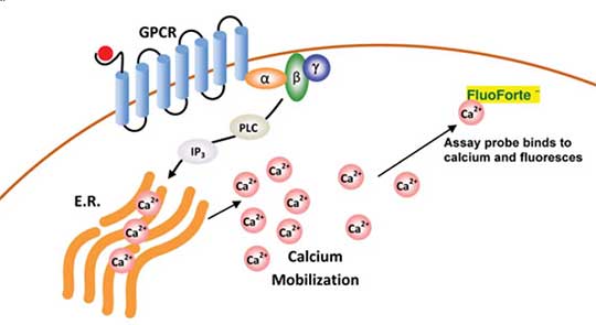
FLUOFORTE® Calcium Assay Kit detects mobilization of intracellular calcium utilizing a fluorogenic calcium-binding dye optimized for superior cell-permeability and retention. The self-quenching dye undergoes an electronic change upon binding of calcium, resulting in a several order of magnitude greater fluorescence.
|

|
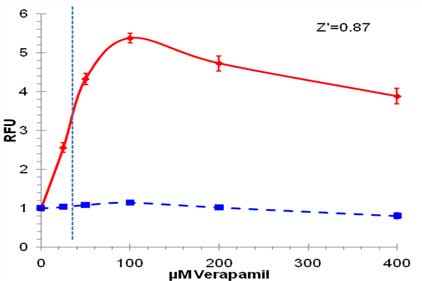
High Throughput Screening of Therapeutics for Lysosome-perturbing Activity using Enzo’s LYSO-ID® Red cytotoxicity kit (GFP-CERTIFIED®) to monitor dysfunction of lysosomal degradation using a drug-like cationic amphiphilic tracer (CAT) dye that rapidly and selectively stains acidic organelles, suitable for monitoring accumulation of lysosomes and lysosome-like structures in live cells. Results show toxicity of Verapamil in U-2 OS cells estimated using a conventional fluorescence microplate reader. U-2 OS cells were treated with Verapamil for 18 hours and stained with LYSO-ID® Red dye for 15 minutes. The high Z-factor (0.87 for 100 µM Verapamil) indicates that LYSO-ID® Red dye is suitable for HTS applications.
|
Cell Source and Types
Cells used in CBAs express the necessary factors and signaling intermediates. Various types and sources of cells have been used. Immortalized cell lines are widely used for drug screening assays because they are cheap, easy to grow, reliable and reproducible. For example, HEK293 cells are an easy, amenable experimental system. However, immortalized cell lines, derived from a well-known tissue type, have undergone significant mutations and their biological characteristics might be altered in the immortalization process and different from those of the native or normal cells. Primary cells may provide representative responses; however, they have a limited life span in culture and are difficult to grow.
Human cancer cell lines represent the cancer of origin and are widely used for anticancer drug screening research. However, they can contain mutations that might affect the experimental results. For instance, the breast cancer cell line MCF-7 lacks a functional caspase-3 gene product. Furthermore, CBAs employing primary cells or immortalized cancer cell lines have been insufficient in developing effective therapeutics for cancers. Recent advances in stem cell research have revolutionized the drug discovery process. Cancer stem cells genetically arise from oncogenic transformation of either stem cells or progenitor cells, can be isolated from tumors and used as an effective platform for cancer drug screening. Embryonic stem cells (ESCs) and induced pluripotent stem cells (iPSCs) can provide an unlimited source of normal human cells that can be used for drug screening and toxicological studies. Recent developments of iPSCs has revolutionized the stem cell field. iPSCs are pluripotent cells artificially derived from somatic cells (e.g., fibroblasts and other adult cell types) by inducing a small set of powerful pluripotency genes. iPSCs can be derived from patients with specific diseases, they have been considered as a new tool in drug discovery. Enzo offers an extensive collection of small molecules and proteins such as growth factors and cytokines that can be used to influence differentiation, biochemical activity, and cell growth.
Cell Culture Systems
Cell culture modes include single cells, monolayer cells on the bottom of the well of a microtiter plate on a two-dimensional (2D) surface. Two-dimensional assays are informative for reporting on a compound’s cell permeability or toxicity. However, they fall short in recapitulating the complex microenvironment and cell-cell interactions of a tissue. Cells cultured in third dimension (3D) scaffolds show similar in vivo morphology with intimate cell-cell and cell extracellular matrix interactions, which are absent in 2D cultures. In 3D cell cultures, cells adhere and grow and maintain the interconnectivity within the 3D constructs. This allows nutrients and metabolites to be transported in and out of the engineered tissues. A wide range of 3D cellular modules have been developed for specific applications such as toxicology, cancer and stem cell drug discovery. The most popular and commonly employed 3D models are single-cell type and mixed co-culture spheroids. A number of assay readouts and detection methods have been applied to 3D cell cultures. Real time analysis have been developed with 3D spheroid model for homogeneous cytotoxicity and hypoxia induction as fluorescence end point read outs. Having the right model for drug screening is essential for predicting which compounds may cause drug-induced liver injury
A broad variety of advanced disease relevant cellular models have been developed using the tools of molecular biology. Advances in micro technologies have led to the development of novel cell-based disease models, micro printed tumor spheroids, tissue-on-a-chip and multicellular organoids. All these innovations offers great hope for new generation of therapies to be discovered. From our long-standing, flagship SCREEN-WELL® Compound library product family to our CELLESTIAL® portfolio of fluorescent probes and assay kits for cellular analysis, Enzo Life Sciences provides a wide variety of products for your drug discovery research needs. Our decades of experience in the design and manufacture of active enzymes and their substrates supports development of an ever-expanding portfolio of biochemical assays. Please check out our Successful Research Tips or feel free to contact our Technical Support Service for further assistance.


