Cytogenetics is the analysis of structure, organization and copy number of chromosomal material and how alterations in the anatomy of chromosomal DNA reflect on biological functions. This branch of genetics focuses on how structural alterations of chromosomes relate to human disease. Cytogenetics tools have driven a vital subset of both research and clinical diagnostics for decades, especially in the fields of obstetrics, gynecology, and oncology. Chromosome alterations form the basis for many diseases affecting patients – from early prenatal defects to cancer. These changes in chromosome structure and ultimately, function, can vary drastically in scale, from absence, fusion or excess of entire chromosomes to alterations at the level of individual genes and even single nucleotide polymorphism (SNP). To detect chromosomal abnormalities, researchers and clinicians can rely on numerous methods to analyze genomic alterations, but depending on the scale and nature of these alterations, one or the other method might be beneficial. And at times, a combination of these methods might be best suited for analysis. Therefore, the analytical weapon of choice should be chosen wisely.
Karyotyping
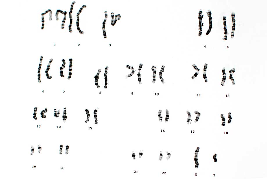
Being one of the oldest genetic methods at a researcher’s disposal, karyotyping predates even our understanding of how genetic information is encoded into the DNA by half a century. A karyotype describes the number and appearance of chromosomes. Normally, karyotyping is performed on spreads of dividing cells to obtain a karyogram (or idiogram), a depiction of condensed metaphase chromosomes rearranged in matching pairs by size. This rearranged photomicrograph is then used to analyze the phenotypic, microscopic appearance of somatic chromosomes as they appear during the mitotic division of a cell. This method allows for efficient and intuitive analysis of chromosomal alterations at the microscopic level with relatively simple means. Typically, this method is improved by additional staining, such as trypsin combined with Giemsa stain in a method called G-banding, because it will stain the chromosomes in typical, reproducible bands. These bands have been for decades the best means to describe chromosome locations and form the basis of modern denomination of chromosomal loci. For example, a gene located on 3p22.1 means that it can be found on the p-arm –the shorter arm, with q being the longer arm – of chromosome 3, in region 2, band 2, sub-band 1. These regions and bands used to describe gene loci originate from defined G-banding of karyotypes.
Alternative staining methods to produce band patterns on metaphase chromosomes are R-banding (i.e. a reverse- G-banding), Q-banding with the use of fluorescent quinarcine, or multicolor FISH banding with the help of a mix of defined, chromosome-specific
fluorescence in-situ hybridization (FISH) probes. Multicolor FISH (or chromosome painting) allows for easier, more instinctive analysis of a karyotype by using a defined mixture of fluorescence probes. Each chromosome is labeled in a unique color, not only allowing to identify chromosomes very easily, but also directly highlighting translocations, where parts of a given chromosome are wrongly placed on another.
Karyotyping in combination with G banding typically offers a spatial resolution to detect chromosomal alterations bigger than 5-10Mbp. It is a cost-effective, methodologically simple screening method for larger cytogenetic alterations and can detect aneuploidy (abnormal numbers of chromosomes or chromosomal regions) as well as transpositions, deletions, duplications, and even inversions of chromosomal arms or larger chromosomal fragments. Typical examples of genetic abnormalities detected by karyotyping include Trisomy 21, Turner Syndrome (only one X chromosome, no Y chromosome) or Cri du Chat (truncated p-arm of chromosome 5). In contrast, Prader-Willi-Syndrome, a genetic disorder where ~75% of the patients show a deletion of the region 15q11-13, is at the detection limit of karyotyping and cannot be reliably diagnosed with this method.
Additionally, karyotyping relies on the availability of fresh, proliferative cell samples. Sampled cells are grown in culture and then arrested in metaphase by the use of
colchicine, which blocks the formation of the mitotic spindle and allows efficient visualization of condensed metaphase chromosomes. This culture method is time-consuming and cannot be performed on growth-arrested or fixed tissue. Imaging and rearrangement of images is not only time-consuming, but also requires experienced personnel to obtain and interpret the karyograms. Nonetheless, karyotyping is a robust and highly useful diagnostic tool that is not only applied in obstetrics and gynecology, but also in cancer medicine.
Fluorescence In Situ Hybridization
FISH as a cytogenetics tool offers much more than chromosome painting for spectral karyotyping. It is a powerful tool to detect and visualize known cytogenetic alterations and is frequently used as a diagnostic means. Utilizing fluorescent FISH probes against known alterations of sequences or viral sequences integrated into the host genome allows for rapid detection of common genetic alterations. Furthermore, multiplexing with several different probes labeled with different fluorophores can easily be achieved. Probes can be purchased ready-to-use or synthesized by techniques such as nick translation using Enzo’s
Nick Translation DNA labeling system 2.0 kit in combination with our
SEEBRIGHT® fluorescent dye-dUTPs. This is a simple, rapid, reliable, and efficient method to generate custom labeled DNA probes for FISH in just one hour. However, FISH as a cytogenetic diagnosis technique is more often performed with ready-made probes against known common and uncommon genomic alterations. FISH’s advantage over karyotyping is that it can be performed in interphase nuclei as well as on fixed samples, thus eliminating the need for upstream cell culture to generate test samples.

Figure 1: Hybridization of a digoxigenin-labeled HPV Type 6/11 probe to cervical biopsy tissue. Visualized with anti-dig-AP and HIGHDEF® red IHC chromogen (AP).
|
Although karyotyping widely remains the method of choice, FISH probes against common aneuploidies are commonly used as a collectively tool called rapid aneuploidy detection (RAD). RAD is used to detect known, mostly common alterations such as insertions, duplications, deletions, translocations, and gene fusions. Especially in cancer oncology research, FISH (or in cases of tissue samples, often chromogenic ISH) is a frequent method of choice to detect common alterations such as deletion of p16 in malignant mesothelioma or insertion of human papilloma virus (HPV) into the genome. An example of this is Enzo’s
PATHO-GENE® virus detection assays.
FISH allows for the rapid and easy detection of known common and uncommon alterations with standard laboratory methods. Additionally, FISH offers a resolution of alterations at the level of a few base pairs, although only within the targeted sequences. It has very limiting sample depth, allowing only for analysis of a few alterations at a time. Furthermore, it is severely hampered by being limited to known and targeted sequence alterations and does not allow for detecting random or unknown alterations. It is thus a diagnostic tool mainly applied for confirmation of suspected diagnoses.
Quantitative Fluorescence Polymerase Chain Reaction
An alternative to FISH-based RAD is Quantitative Fluorescence Polymerase Chain Reaction (QF-PCR). This method utilizes fluorescence-labeled DNA primers against chromosome-specific repeat sequences and PCR to amplify the target sequences. Differences in the number of repeats per allele will result in different length of the amplified products. Size and fluorescence intensity of the amplified segments is quantified and summarized in a graphic representation which allows for detection of copies of different alleles as well as duplication of identical alleles. However, the results will be uninformative, should the repeat sequences be homozygous and identical for all alleles present in the sample. Like FISH, QF-PCR is limited to known and targeted alterations only. However, relying on DNA samples, it is generally easier to obtain sample templates and the technical and equipment requirements are relatively simple. It is commonly used to detect trisomies 13, 18, 21, and abnormalities of the sex chromosomes. Unfortunately, initial installation costs have to be overcome with high sample numbers and frequent analyses. However, running costs are relatively low and results are easy to interpret.
Comparative Genomic Hybridization
Originally developed for the evaluation of differences between solid tumor genome and normal tissue, comparative genomic hybridization (CGH) is based on competitive binding of two DNA samples to a chromosome-wide selection of DNA templates. In CGH, two sets of whole genome DNA preparations, a healthy reference standard and the test sample, are used to generate fluorescent hybridization probes in two different colors, most commonly red and green. At Enzo, we offer excellent
labeling solutions for your CGH hybridization probes. These probes are hybridized to a genome-wide template and genomic regions present in both test sample and reference standard appear yellow, while regions over or underrepresented in the test sample appear red or green. The original method of using normal metaphase spreads of chromosomes similar to karyotyping as a template to hybridize to, has meanwhile fallen into complete disregard and is widely replaced by array CGH (aCGH). Here, oligonucleotides generated from chromosomal DNA, bacterial artificial chromosomes (BACs) or by oligo-synthesis are spotted or printed to a glass slide and serve as a hybridization template to generate a microchip array. Chip-based aCGH allows for the automated detection of hundreds of thousands of known target sequences. Whole genome arrays consist of target sequences that offer genome-wide, extensive, and in regions of specific interest, often continuous coverage. Targeted arrays, in contrast, target specific regions of interest only, but offer increased detail and redundancy to improve confidence in the result. Results are comprised in a “virtual karyotype” – a graphic representation of all hybridization probes sorted in chromosomal order, and can be used to detect copy number variations (CNVs). Resolution of genome-wide CGH arrays commonly reaches 50-80Kbp and below, with targeted arrays being able to offer a fraction of this resolution. Resolution and resulting versatility in detecting CNVs make aCGH the gold standard of cytogenetics.
Array-CGH can also detect mosaicism, where a sample contains a mixture of cells with different copy numbers of alleles, as long as the sample is present in substantial amounts. Mosaicism can often be found in tumor samples that are mixed with normal cells, as well as in some genetic disorders.

Figure 2: A virtual karyotype obtained by aCGH. Reference male DNA was compared to sample DNA using a 1x1M Human CGH SurePrint array from Agilent in combination with Enzo’s CYTAG® CGH labeling kit. Karyotype shows a healthy male subject.
|
A limiting disadvantage of CGH is its inability to detect copy number neutral aberrations such as balanced chromosomal translocations and inversions. This limit can partially be overcome by complementing aCGH with array probes for known single nucleotide polymorphisms (SNPs), known allele variants that differ –often inconsequential– in a single base pair. Sequences for known variants of SNPs are spotted additionally to standard CGH probes onto the array and allow for targeted detection of specific allele variants. Thus, CGH+SNP arrays allow detection of copy-neutral haplotype variations and genomic aberrations like uniparental isodisomies, where both copies of one allele are identical due to deletion of one parental allele and replacement by the other through duplication, resulting in copy-neutral loss of heterozygosity. As this type of genetic aberration cannot be detected by conventional cytogenetics, including aCGH, SNP-based arrays are preferred for virtual karyotyping of tumors. Array-CGH+SNP also allows for determining genetic identity by descent. No matter which application or CGH method is being used, the selection of the right chip requires careful consideration to choose the right assay.
Next-Generation Sequencing
Massive parallel sequencing or next generation sequencing (NGS) is becoming a ubiquitous technology in basic biology research and starting to make its way into both diagnostic and clinical settings. While the technologies collectively known as NGS vary greatly, they are all basically more robust versions of classical Sanger sequencing. Similar to Sanger sequencing, small DNA fragments are sequenced linearly, but millions of sequences are obtained in parallel in a fully automated manner by high-throughput data acquisition. Each base pair is sequenced multiple times for accuracy. NGS allows for sequencing of the entire genome within as little as one day and thus offers a formidable tool for genome-wide individual analyses.
The process can be streamlined and sped up further if sequencing is focused on specific areas of interest only, such as certain chromosomes or regions. A common approach is to sequence only the ~22-thousand coding genes in a process called whole exome sequencing. Another method is to focus on relevant target genes only, like known oncogenes in tumor analysis. NGS is a powerful cytogenetic tool that allows detection of all types of cytogenetic aberrations, from common insertions or deletions to mosaicism and single nucleotide mutations, and even unknown inserts of pathogen DNA. Furthermore, it is extremely sensitive, so that even detection of fetal DNA from maternal blood becomes possible, and risky procedures to obtain amniotic fluid or placental cells can be omitted. These benefits make NGS an extremely powerful and attractive tool.
Although costs per reaction are relatively low, clinical application of NGS is not cost effective because it requires a high frequency of analysis and initial setup costs for obtaining the necessary machinery and infrastructure are substantial. Additionally, the requirement for experienced personnel due to the vast amount of data generated, the necessary skillful analysis, and interpretation of unknown genetic variants are an additional and lengthy caveat. Therefore, successful implementation of NGS as the cytogenetic gold standard might not be possible without supra-regional centralization.
Applications of Different Cytogenetics Methods
Due to the differences in resolution and the various benefits and limitations of each technique, great care should be taken when deciding which tests to perform. Additionally, interpretation of the results requires skillful analysis, as there might be a discordance between different methods for certain specific findings. Karyotyping remains the method of choice for common aneuploidy assessment, as in the analysis of indeterminate gender or fertility issues. FISH is a powerful tool for confirmation of well-known genetic abnormalities, as well as the analysis of oncogenetic aberrations and detection of known pathogens, and the only cytogenetics technique applicable to fixed samples. CGH and SNP is the method of choice for detection of more complex cytogenetic aberrations to explain learning difficulties, intellectual disabilities, developmental delay, behavioral problems including autism, or miscarriages. NGS offers the most comprehensive cytogenetics analysis but its limitations due to its complexity will confine it for the time being to analysis of complex cases and research settings.
Especially in cancer research, a single cytogenetics technique might not generate the needed clarity of results for clinical decisions, and cytogenetics research may continue relying on a combination of different techniques to obtain and confirm results, including techniques at the fringes between cytogenetics and molecular biology, such as chromosome conformation capture, chromatin immunoprecipitation, and others. Ultimately, individual cytogenetics techniques have their strengths and weaknesses, and the choice of the right test might further vary depending on available expertise and familiarity of the user with the methodology.
Enzo Life Sciences is a global leader in DNA and RNA labeling technologies. We offer a range of products for your
Genomics and
Cancer research needs. Our
Array CGH labeling solutions offer outstanding label incorporation and unrivaled DLR scores. For a simple and efficient method for generating labeled DNA, please check out our
nick translation DNA labeling kit as well as a list of our
SEEBRIGHT® fluorescent dye-dUTPs. While you are at it, please check out our TechNotes on
tips for successful aCGH and
FISH. For all questions and concerns regarding any of our products, our
Technical Support Team is here to assist.