Dopamine is an organic chemical of the catecholamine and phenethylamine families. Dopamine functions as a neurotransmitter in the brain.
Dopaminergic signaling is associated with reward-motivated behavior and motor control with dysfunction of the dopamine system leading to numerous diseases. For example, degenerative Parkinson’s disease is caused by the loss of dopamine-secreting neurons that leads to motor impairment. Enzo Life Sciences offers a
Dopamine ELISA Kit for the quantitative measurement of dopamine concentration in serum, plasma, and cell culture supernatants.
Initial Discovery of Dopamine’s Role as a Neurotransmitter
Before 1957, the prevailing viewpoint was that 3-hydroxytyramine was an intermediate in the synthesis of norepinephrine and adrenaline from tyrosine. However, between 1957 and 1959, parallel efforts by Kathleen Montagu and her colleagues at Hans Weil-Malherbe lab at Runwell Hospital (England) and Arvin Carlsson and his colleagues at Lund University (Sweden) helped lead to the initial findings that would collectively suggest dopamine’s role as a neurotransmitter in the human brain. In August 1957, Montagu published the first paper that demonstrated her findings on key neurotransmitters. As part of her research, she ran a column-based assay to examine the quantities of noradrenaline, adrenaline, and 3-hydroxytyramine from extracted tissues of the brains of several species (rat, rabbit, guinea pig, chick, human, and frog). Montagu speculated that there may be an additional catecholamine similar to hydroxytyramine, which she later confirmed to be 3,4-dihydroxytyramine (“dopamine”) via paper chromatography of the eluates from the resins she used for her extracted brain tissues. In November 1957, Carlsson found that he could reverse the akinetic effects that reserpine induced in his rabbits by injection of dopamine and norepinephrine precursor, 3, 4-dihydroxyphenylalanine (L-DOPA) intravenously and found that it correlated to a recovery of dopamine but not noradrenaline. This data suggested that the lack of dopamine may have been responsible for the akinetic state seen in his animals. Eventually, Carlsson’s group developed an assay that could measure dopamine concentration in the brain and mapped out where the highest concentration of dopamine was present. They determined that dopamine was found in high concentrations in the striatum, the largest component of the basal ganglia. At the time, it was already well-known that the basal ganglia played a key role in voluntary motor functions. These findings helped to shape the initial hypotheses that dopamine may be a key neurotransmitter in the control of motor function.
What is Dopamine? Where is it produced in the brain? How is it produced?
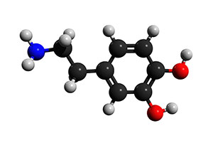 Figure 1:
 Dopamine neurotransmitter molecule
Since its initial discovery, Dopamine, also known as 3, 4-Dihydroxytyramine, has been heavily characterized. It is made up of a benzene ring with two hydroxyl side groups attached to one amine group via an ethyl group. It is produced by dopaminergic neurons in the brain from tyrosine via the addition of a hydroxyl group which transforms it to L-DOPA (or Levo-DOPA) and subsequent removal of a carboxylic acid group from the ethyl side chain linked to the amine group resulting in Dopamine. The dopaminergic neurons that produce this signaling molecule are located in the brain at the substantia nigra and ventral tegmental area which are both located in the midbrain as well as the arcuate nucleus of the hypothalamus. Dopamine serves as a neurotransmitter—a chemical released by neurons to transmit an electrical signal chemically between one neuron to the next to pass on a signal to and from the central nervous system. After production of dopamine, the neurotransmitter is packaged into a synaptic vesicle, vesicular monoamine transporter 2 (VMAT2) and stored until action potentials induce the release of dopamine into the synaptic cleft and cause binding to dopamine receptors on the postsynaptic neuron.
Dopamine neurotransmitters bind to five subtypes of dopamine receptors: D1, D2, D3, D4, and D5, which are members of the G-protein coupled receptor family that are divided into two major subclasses: D-1-like and D-2-like. The binding of dopamine to these receptors initiates cascades of signaling responsible for activating functions in the associated areas of the brain where each receptor type is most prevalent. D1-like receptors are more prevalent than D2-like receptors. To understand how dopamine functions in the human brain as a neurotransmitter requires looking at the effect of dopamine binding to D1-like and D2-like receptor types to exert their effects via second messenger systems. The binding of dopamine to D1-like receptors (D1 and D5) results in excitation via the opening of Na+ channels or inhibition via the opening of K+ channels. D1-like receptor stimulation induces the activation of adenylate cyclase, the enzyme that converts ATP to cAMP, thereby increasing cAMP levels leading to the disinhibition of protein kinase A (PKA) which phosphorylates downstream targets such as cAMP regulatory element binding protein (CREB). The translocation of CREB to the nucleus and CREB-dependent transcription of numerous genes is responsible for the synaptic plasticity necessary for learning and memory formation. In contrast, D-2 like receptor binding (D2, D3, and D4) lead to inhibition of the target neuron by exerting an opposite effect of inhibiting adenylate cyclase through coupling to G proteins Gi/o which decreases the production of cAMP. Whether dopamine is excitatory or inhibitory is a matter of which type of effect on a target neuron is exerted which is based on which types of receptors are on the membrane surface of the neuron and how the neuron responds to increases or decreases in cAMP concentration.
What does Dopamine do in the Human Brain?
Dopamine plays important roles in executive function, motor control, motivation, arousal, reinforcement, and reward through signaling cascades that are exerted via binding to dopaminergic receptors at the projections found in the substantia nigra, ventral tegmental area, and arcuate nucleus of the hypothalamus of the human brain.
In the substantia nigra, the nigro-striatal pathway projects dopaminergic neurons from the input area (known as the pars compacta) to the dorsal striatum and plays a primary role in the control of motor function and learning motor skills. If the dopaminergic neurons in the nigro-striatal pathway degenerate, this causes a dysregulation of motor control, a hallmark of Parkinson’s Disease.
In the ventral tegmental area (VTA), the mesolimbic pathway projects from the prefrontal cortex to the nucleus accumbens of the amygdala, cingulate gyrus, hippocampus, and pyriform complex of the olfactory bulb. The dopaminergic projections in the amygdala and cingulate gyrus are responsible for emotion formation and processing. In the hippocampus, the presence of dopaminergic neurons is associated with learning, working memory, and long-term memory formation. Lastly, the pyriform complex of the olfactory bulb is responsible for providing humans with the sense of smell. In the mesolimbic pathway, dopamine is released during pleasurable situations, causing arousal and influences behavior (motivations) to seek out the pleasurable activity or occupation and bind to dopaminergic receptors present in the nucleus accumbens and prefrontal cortex. Increased activity in the projections to the nucleus accumbens play a major role in reinforcement and in more extreme cases with addictions.
In the arcuate nucleus of the hypothalamus, dopamine neurons make up the tuberoinfundibular pathway which projects to the pituitary gland and inhibits the secretion of the hormone prolactin. Dopamine produced by neurons in the arcuate nucleus are released in the hypothalamo-hypophysial blood vessels which supply the pituitary gland with dopamine to inhibit the production of prolactin.
Quantitative Measurement of Dopamine Levels in Samples
Assessing dopamine levels as they pertain to de-regulation of functions associated with certain portions of the brain is an attractive target for neuroscience research. Enzo Life Sciences offers a
Dopamine ELISA Kit, which is a colorimetric competitive immunoassay capable of quantifying dopamine in serum, plasma, tissue homogenates and other biological fluids. This kit is highly specific for human dopamine and has negligible cross-reactivity between detection of human dopamine and its analogues. This highly sensitive immunoassay has a detection range that can detect as little as 1.56 ng/ml and as much as 100ng/ml (Figure 1). Furthermore, this product was designed with high sensitivity, high lot-to-lot reproducibility, and low time-to-result and offers a simple protocol that can produce reliable, quantitative results for our end-users in less than 2 hours for up to 40 samples in duplicate.
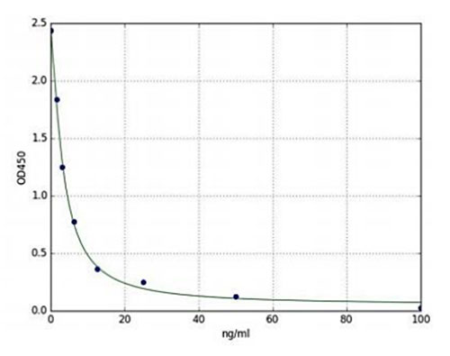
Figure 2: Standard curve of the Dopamine ELISA Kit (ENZ-KIT188) depicting a typical standard curve (1.56 ng/ml-100ng/ml).
|
Enzo Life Sciences provides a wide variety of products for your
Neuroscience and
Immunology research needs. We offer other neurotransmitter immunoassays such as our
Serotonin ELISA Kit and
Histamine ELISA Kit and a large selection of antibodies to study neurotransmitters such as ACTH, ANP, BNP, CCK, CGRP, NPY, GABA, GLP-1, and Substance P. Enzo’s comprehensive portfolio includes our
SCREEN-WELL Neurotransmitter Library, which contains 661 CND receptor ligands in a 96-well format. Please check out our Neuroscience and
Cell Signaling/Signal Transduction platforms for more information or feel free to contact our
Technical Support Service for further assistance.