A tumor or a neoplasm is the result of an excessive and uncontrolled proliferation of a single normal cell, which has been transformed into a cancerous state following multiple cellular alterations and rendered resistant to apoptosis, cell-to-cell contact inhibition, growth factor removal or immune cells. There are now compelling evidences that both solid tumors and hematological malignancies are in fact very heterogeneous by nature. They are made of different populations of cells, notably one that possesses the stem cell properties of self-renewal and differentiation. These cells are referred to as cancer stem cells or cancer-initiating cells. It is a clearly distinct subpopulation within the tumor and they are thought to be responsible for the recurrence of cancer and the spreading of cancer cells to other parts of the body via the process of metastasis. Like normal stomatic stem cells, cancer-initiating cells are resistant to chemotherapeutic agents allowing them to eventually re-populate the tumor site. They have a slow rate of cell turnover making them less responsive to chemotherapeutic agents and express different efflux pumps or transporters from the ABC family capable of pumping out the drugs. Based on diametrically opposite observations made in pancreatic and breast cancers, it is now hypothesized that cancer stem cells are made of stationary cancer stem cells that persevere in differentiated areas as well as mobile cancer stem cells localized near the tumor surface which are capable, upon epithelial-to-mesenchymal transition, of invading and metastasizing distant sites.
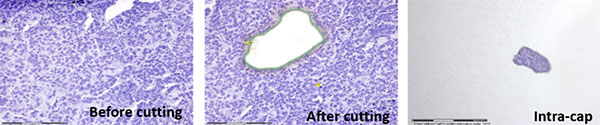
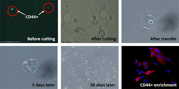
Despite a large number of studies on the subject of cancer stem cells, very little is known about their genomic, transcriptomic and proteomic signatures. For a very long time, tumors used to be studied as a whole. As a consequence of this heterogeneity, DNA, RNA or protein contents were considerably diluted out and their analysis did not provide an accurate picture. A critical breakthrough came from the development of laser capture microdissection (LCM), a technique allowing for the isolation of foci of tumor cells within a heterogeneous tissue (Fig. 1) and the enrichment of specific cells in culture (Fig. 2). A brief laser pulse is directed towards a specific area to specifically select and isolate cancer cells for further genetic or proteomic analysis. Combining LCM with transcriptomic approaches opened the way to precision cancer biology and several key diagnostic and prognostic biomarkers as well as therapeutic targets were identified in the process.
Figure 1: Laser capture microdissection of cells from cancer biopsies.
Figure 2: Laser capture microdissection and enrichment of CD44+ breast cancer cells.
More recently, Adam Marcus and Jessica Konen from Emory’s Winship Cancer Institute extended that approach of precision cancer biology to not only highlight the heterogeneity of a tumor but also track, isolate and understand cancer cells that may behave differently from the pack. Metastasis is the main cause of lethality in cancer patients but not all cancer cells have the ability of metastasizing. The idea was therefore to isolate cells that do, those so-called mobile cancer stem cells. Cancer cells were labeled with a green-to-red photo-switchable fluorescent marker. Cells with an ability to migrate away from 3-D spheroid colonies were selected and in response to an intense laser exposure, photo-converted from green to red. Cancer cells labeled in red can then be tracked for further observations about their phenotypical behavior or alternatively, sorted out for subsequent gene expression microarray analysis or mass spectrometry-based proteomic study. The scientists’ technique was termed spatiotemporal genomic analysis or SAGA and should help researchers answer questions about migration, migratory cells and metastasis. In other words, are migratory cells actual mobile cancer stem cells and if so, do they need the help of other cells to form a metastasis? More importantly, researchers may have a better understanding of cancer cell biology as a whole thanks to this approach and hopefully, make significant progress towards therapies to impede metastasis.
Video: Chasing metastatic cancer cells
Enzo Life Sciences offers a comprehensive portfolio for cancer research and stem cell discovery including cell migration assays, antibodies and immunoassays against invasion markers, and live cell analysis tools, some of which are described below.
Ultra-sensitive ELISA kit for the quantitative detection of human VEGF with 100% cross reactivity to VEGF 165 and negligible cross-reactivity to VEGF 121 and an assay time of just 2.5 hours