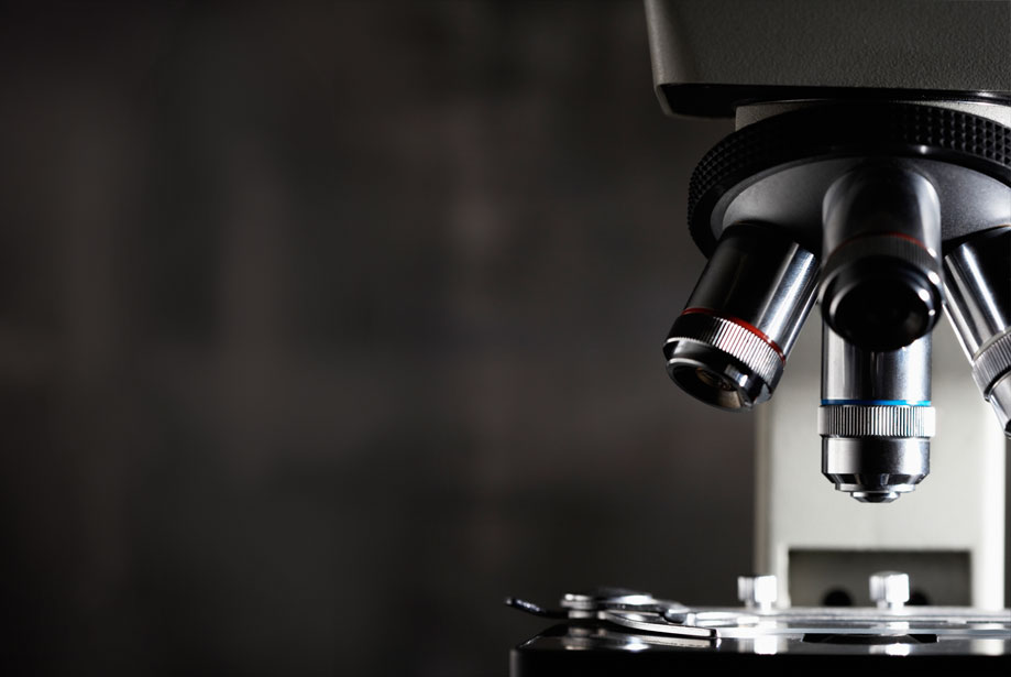
Immunohistochemistry is a widely used biological technique allowing the end user not only to analyze the anatomy of the tissue of interest but also to visualize the distribution, the localization and the intensity of the expression of a specific antigen or cellular components in tissue sections.
It can be summarized in three major steps: (1) the binding of the primary antibody to a specific antigen, (2) the formation of an antibody-antigen complex following the addition of a secondary enzyme-conjugated detection antibody and finally, (3) the presence of a chromogenic substrate which in contact of the enzyme will lead to the generation of a colored deposit at the site of the antibody-antigen complex.
Nowadays, the efficient diagnosis of a cancer as benign or malignant and the determination of the stage and grade of a cancer rely heavily on the expression analysis of tumor biomarkers by immunohistochemistry.
There are now several biomarkers that are validated for diagnostic use with different cancers featured with different panels of biomarkers.
However, there is an urgent requirement for new prognostic markers.
Despite the recent surge of new technologies such as Next-Generation sequencing or Imaging MALDI mass spectrometry which monitor the expression of new predictive targets, researchers and clinicians still rely heavily on the use of immunohistochemistry.
Recently, Dr. Ohnishi and coworkers from Kumamoto University hypothesized that a sialic acid receptor (CD169) that is expressed on lymph node sinus macrophages could have significance in predicting prognosis for patients with colorectal carcinoma (CRC).
They first demonstrated that CD169 expression was up-regulated in macrophages stimulated with type I interferons in vitro.
Using immunohistochemistry to look at the expression of CD169 in samples of regional lymph nodes, they noticed an inverse correlation between the density of CD169-positive cells and tumor enlargement or lymph node metastasis.
The latter is generally associated with poor prognosis in CRC patients.
However, when studying the clinicopathological features of 83 CRC patients, a positive correlation between high density of CD169-positive cells in regional lymph nodes and overall survival was observed.
The common consensus in tumor immunity is that the presence of macrophages mediates the activation of CD8-positive cytotoxic T cells.
Using three-color immunostaining, they showed that CD169, CD8 and CD43 (i.e. a CD169 ligand found on the surface of CD8+ T cells) were co-expressed in regional lymph nodes of CRC patients and that CD169-positive macrophages directly interacted with CD8- and CD43-positive cytotoxic T cells in regional lymph nodes.
Finally, they demonstrated that patients with a high density of CD169-positive cells also had high levels of infiltration of CD8-positive T cells in their tumor tissues.
This study suggests that the number of CD169-positive cells in regional lymph nodes improves prognosis by enhancing cytotoxic immune response against the tumor and consequently, is beneficial for overall survival.
It also emphasizes the importance of immunohistochemistry, not only in diagnosis, but also in prognosis.
This demonstrates the added value in monitoring the expression of a panel of biomarkers and the interaction of several populations of cells on single tissue sections.
Enzo Life Sciences offers histopathologists a comprehensive portfolio for immunohistochemistry including antibodies, tissue microarray, retrieval reagents, detection reagents and a wide variety of chromogens allowing multi-color immunohistochemistry staining, some of which are described below: