| Alternative Name: | Lymphocyte activation gene-3, FDC protein, CD223 |
| |
| Clone: | L4-PL33 |
| |
| Host: | Mouse |
| |
| Isotype: | IgG1κ |
| |
| Immunogen: | Synthetic peptide corresponding to 30 aa (GPPAAAPGHPLAPGPHPAAPSSWGPRPRRY) from the first N-terminal D1 domain of human LAG-3 (lymphocyte activation gene-3). |
| |
| UniProt ID: | P18627 |
| |
| Source: | Purified from tissue culture supernatant. |
| |
| Species reactivity: | Human
|
| |
| Specificity: | Recognizes the 30 aa extra-loop of the first N-terminal D1 domain of LAG-3. |
| |
| Applications: | ELISA, Flow Cytometry, ICC, IHC, IP, WB
|
| |
| Recommended Dilutions/Conditions: | Flow Cytometry (1:100)
Immunohistochemistry (frozen sections, 1:150, FFPE sections, 1:150)
Western Blot (1-5 µg/ml)
Immunoprecipitation (10 µg/ml)
Suggested dilutions/conditions may not be available for all applications.
Optimal conditions must be determined individually for each application. |
| |
| Purity Detail: | Protein A affinity purified. |
| |
| Formulation: | Liquid. In PBS containing 10% glycerol. |
| |
| Use/Stability: | Stable for at least 6 months after receipt when stored as recommended. |
| |
| Handling: | Avoid freeze/thaw cycles. |
| |
| Shipping: | Blue Ice |
| |
| Short Term Storage: | +4°C |
| |
| Long Term Storage: | -20°C |
| |
| Scientific Background: | The lymphocyte activation gene-3 (LAG-3, CD223), a member of the immunoglobulin superfamily (IgSF) related to CD4, binds to the major histocompatibility complex (MHC) class II molecules but with higher affinity than CD4. Several alternative mRNA splice-variants of human LAG-3 have been described, two of them encoding potential secreted forms: LAG-3V1 (i.e. the D1-D2 domains of the protein, 36 kDa) and LAG-3V3 (D1-D3, 52 kDa). The longer form was detected by ELISA in the serum of healthy individuals as well as of tuberculosis patients with a favorable outcome. LAG-3 expression by T cell clones correlated with IFN-γ production, and hence soluble LAG-3 has been suggested as a serological marker of Th1 responses. |
| |
| Regulatory Status: | RUO - Research Use Only |
| |
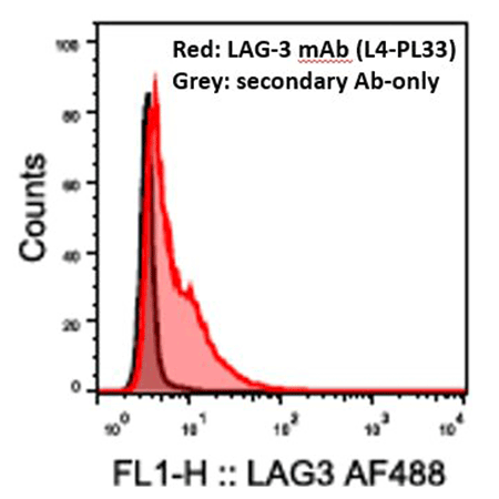
Flow cytometry analysis of 0.5x10^6 THP-1 cells stained using LAG-3 mAb (L4-PL33) at a concentration of 5μg/ml, followed by Alexa Fluor® 488 conjugated anti-mouse secondary antibody.
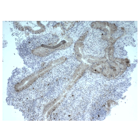
Immunohistochemistry analysis of formalin-fixed, paraffin-embedded human tonsil tissue stained with LAG-3 (human) recombinant mAb (L4-PL33) at 6μg/ml.
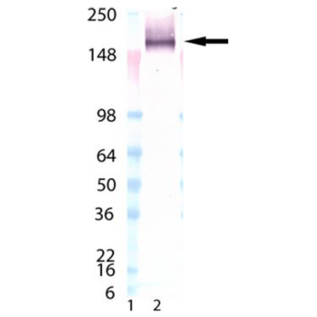
Western blot analysis of LAG-3 mAb (L4-PL33) (Prod. No. ENZ-ABS677): Lane 1: MW marker, Lane 2: LAG-3 (human):Fc (human), (recombinant) (Prod. No. ALX-522-078).


