-
Far-red fluorescent specific DNA dye
-
Stable and highly pure
-
Live, permeable and fixed cells can be analyzed
-
No photobleaching effect
-
No RNase treatment is required
-
GFP and FITC compatible
-
UV laser source is not required for excitation
-
Validated for a wide range of cell densitites
-
Quick and easy to use!
The NUCLEAR-ID® Red DNA Stain is a cell permeable dye, designed for use in a range of fluorescence detection technologies, in the discrimination of nucleated cells. It is resistant to photobleaching and is suitable for live-cell staining of nuclei. Also this dye provides a convenient approach for studying the induction and inhibition of cell cycle progression by flow cytometry. Potential applications of this reagent for live-cell studies are in the determination of cellular DNA content and cell cycle distribution, for the detection of variations in growth patterns, for monitoring apoptosis, and for evaluating tumor cell behavior and suppressor gene mechanisms.
Product Details
| Quantity: | 200µl |
| |
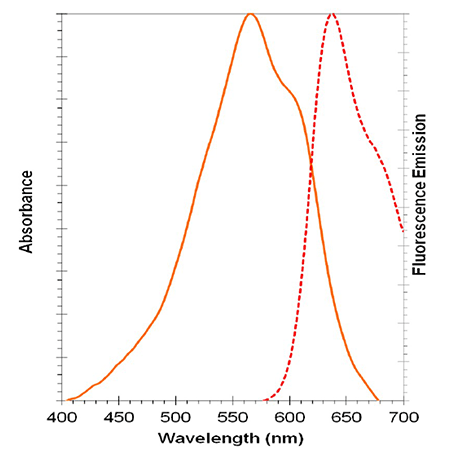
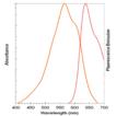
| Excitation maximum: | 566nm |
| |
| Emission maximum: | 650nm |
| |
| Purity: | ≥93% (HPLC) |
| |
| Quality Control: | - Absorption peak of NUCLEAR-ID® Red dye: λmax = 566 ± 4 nm
- % purity of NUCLEAR-ID® Red dye by HPLC: ≥93%
|
| |
| Appearance: | Frozen liquid. |
| |
| Applications: | Flow Cytometry, Fluorescent detection
|
| |
| Shipping: | Blue Ice |
| |
| Short Term Storage: | -20°C |
| |
| Long Term Storage: | -80°C |
| |
| Handling: | Protect from light. Avoid freeze/thaw cycles. |
| |
| Technical Info/Product Notes: | The NUCLEAR-ID® Red DNA Stain is a member of the CELLESTIAL® product line, reagents and assay kits comprising fluorescent molecular probes that have been extensively benchmarked for live cell analysis applications. CELLESTIAL® reagents and kits are optimal for use in demanding imaging applications, such as confocal microscopy, flow cytometry and HCS, where consistency and reproducibility are required. |
| |
| Regulatory Status: | RUO - Research Use Only |
| |
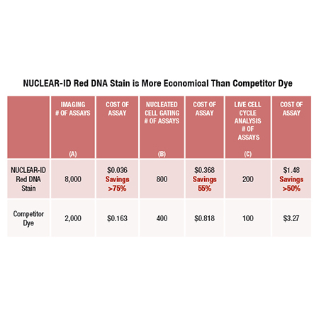
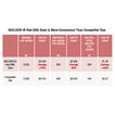
Relative costs of using NUCLEAR-ID Red DNA in comparison to competitor dye in various applications: (A) Imaging (visualization), (B) Nucleated Cell Gating (flow cytometry) and (C) Live Cell Cycle analysis using flow cytometry. Dilutions can vary depending on cell strain and cell concentration.Notes:Assumes staining of a 100 µL staining volumeAssumes staining of a 500 µL cell suspension volumeAssumes a staining of 500 µL cell suspension volume
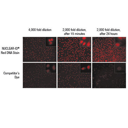
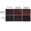
NUCLEAR-ID Red DNA Stain requires lower concentration than competitor’s dye to visualize dsDNA.HeLa cells were grown to ~60% confluency. Cells were stained with NUCLEAR-ID Red DNA Stain at a final concentration of 4000x or 2000x or with a competitor’s dye at an equivalent µM concentration at 37°C and gently washed post-staining. Cells were imaged at 15 min and 24h. Results show that 4000x NUCLEAR-ID Red DNA Stain was required for visualization of the dsDNA, while equivalent to 2000x was required for the competitor’s dye. At 24h, the competitor’s dye intensity and cell growth were dramatically reduced at the 2000x equivalent final concentration. At the same time point, 2000x of NUCLEAR-ID Red DNA Stain did not affect cell growth or fluorescent intensity. The NUCLEAR-ID Red DNA Stain shows lower cytotoxicity and requires lower concentration in live cell studies, resulting in lower costs.
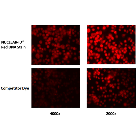
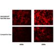
NUCLEAR-ID Red DNA Stain requires lower concentration than competitor’s dye to visualize dsDNA.HeLa cells were grown to ~60% confluency. Cells were stained with NUCLEAR-ID Red DNA Stain at a final concentration of 4000x or 2000x or a competitor’s dye at the equivalent µM concentration at 37°C and gently washed post-staining. Cells were imaged at 15 min.
Please mouse over
Product Literature References
CD200R1L is a functional evolutionary conserved activating receptor in human neutrophils: M.I.P. Ramos, et al.; J. Leukoc. Biol.
111, 367 (2022),
Abstract;
FOXM1 network in association with TREM1 suppression regulates NET formation in diabetic foot ulcers: A.P. Sawaya, et al.; EMBO Rep.
23, e54558 (2022),
Abstract;
Multi-dimensional-double-spiral (MDDS) inertial microfluidic platform for sperm isolation directly from the raw semen sample: H. Jeon, et al.; Sci. Rep.
12, 4212 (2022),
Abstract;
Full Text
Single-cell monitoring of dry mass and dry mass density reveals exocytosis of cellular dry contents in mitosis: T.P. Miettinen, et al.; Elife
11, 76664 (2022),
Abstract;
DYRK1A negatively regulates CDK5-SOX2 pathway and self-renewal of glioblastoma stem cells: B. Chen, et al.; Int. J. Mol. Sci.
22, 4011 (2021),
Abstract;
Full Text
Global phosphoproteomics reveals DYRK1A regulates CDK1 activity in glioblastoma cells: A. Recasens, et al.; Cell Death Discov.
7, 81 (2021),
Abstract;
Full Text
Hydrogen Peroxide Is Crucial for NLRP3 Inflammasome-Mediated IL-1β Production and Cell Death in Pneumococcal Infections of Bronchial Epithelial Cells; Journal of Innate Immunity: S. Surabhi, et al.; J. Innate Immun.
10, 1159 (2021),
Abstract;
Licochalcone A inhibits hypoxia-inducible factor-1α accumulation by suppressing mitochondrial respiration in hypoxic cancer cells: M.K. Park, et al.; Biomed. Pharmacother.
133, 111082 (2021),
Abstract;
NINJ1 mediates plasma membrane rupture during lytic cell death: N. Kayagaki, et al.; Nature
591, 131 (2021),
Abstract;
Overlap of NatA and IAP substrates implicates N-terminal acetylation in protein stabilization: F. Mueller, et al.; Sci. Adv.
7, eabc8590 (2021),
Abstract;
Full Text
The SGLT-2 inhibitor empagliflozin improves myocardial strain, reduces cardiac fibrosis and pro-inflammatory cytokines in non-diabetic mice treated with doxorubicin: V. Quagliariello, et al.; Cardiovasc. Diabetol.
20, 150 (2021),
Abstract;
Full Text
Therapeutic ACPA inhibits NET formation: a potential therapy for neutrophil-mediated inflammatory diseases: R.G.S. Chirivi, et al.; Cell. Mol. Immunol.
18, 1528 (2021),
Abstract;
Full Text
PIE-scope, integrated cryo-correlative light and FIB/SEM microscopy: S. Gorelick, et al.; Elife
8, e45919 (2019),
Abstract;
Full Text
Preclinical evaluation of the pan-fgfr inhibitor ly2874455 in frs2-amplified liposarcoma: R. Hanes, et al.; Cells
8, 189 (2019),
Application(s): IncuCyte® image capture of dedifferentiated liposarcoma cell lines,
Abstract;
Full Text
A high-throughput real-time imaging technique to quantify NETosis and distinguish mechanisms of cell death in human neutrophils: S. Gupta, et al.; J. Immunol.
200, 869 (2018),
Application(s): IncuCyte® image capture of neutrophils,
Abstract;
Full Text
Altered secretion patterns and cell wall organization caused by loss of PodB function in the filamentous fungus Aspergillus nidulans: K.R. Boppidi, et al.; Sci. Rep.
8, 11433 (2018),
Application(s): Fluorescence microscopy of stained fungal mycelia,
Abstract;
Full Text
Caspase-11 auto-proteolysis is crucial for noncanonical inflammasome activation: B.L. Lee, et al.; J. Exp. Med.
215, 2279 (2018),
Application(s): IncuCyte® image capture of macrophages,
Abstract;
Full Text
Fluorescence correlation spectroscopy on genomic DNA in living cells: C. Hodges, et al.; Methods Mol. Biol.
1814, 415 (2018),
Abstract;
ZEB1-repressed microRNAs inhibit autocrine signaling that promotes vascular mimicry of breast cancer cells: E.M. Langer, et al.; Oncogene
37, 1005 (2018),
Abstract;
Full Text
CDK5RAP2 interaction with components of the Hippo signaling pathway may play a role in primary microcephaly: S.K. Sukumaran, et al.; Mol. Genet. Genomics
292, 365 (2017),
Application(s): Flow cytometry using primary fibroblasts,
Abstract;
Full Text
Nesprin-2 Interacts with Condensin Component SMC2: X. Xing, et al.; Int. J. Cell Biol.
2017, 8607532 (2017),
Abstract;
Full Text
Synthesis and biological evaluation of kresoxim-methyl analogues as novel inhibitors of hypoxia-inducible factor (HIF)-1 accumulation in cancer cells: S. Lee, et al.; Bioorg. Med. Chem. Lett.
27, 3026 (2017),
Application(s): Confocal microscopy using HCT116 cells,
Abstract;
How azobenzene photoswitches restore visual responses to the blind retina: I. Tochitsky, et al.; Neuron
92, 100 (2016),
Application(s): Confocal microscopy using retinal ganglion cells,
Abstract;
Full Text
Real-time tracking of parental histones reveals their contribution to chromatin integrity following DNA damage: S. Adam, et al.; Mol. Cell
64, 65 (2016),
Application(s): Confocal microscopy using U2OS cells,
Abstract;
Full Text
Cellular IAP proteins and LUBAC differentially regulate necrosome-associated RIP1 ubiquitination: M.C. de Almagro, et al.; Cell Death Dis.
6, e1800 (2015),
Abstract;
Full Text
Plasminogen activator inhibitor-1 regulates tumor initiating cell properties in head and neck cancers: Y.C. Lee, et al.; Head Neck
38 Suppl 1, E895 (2015),
Abstract;
Pervasive axonal transport deficits in multiple sclerosis models: C.D. Sorbara, et al.; Neuron
84, 1183 (2014),
Abstract;
Zscan4 is regulated by PI3-kinase and DNA-damaging agents and directly interacts with the transcriptional repressors LSD1 and CtBP2 in mouse embryonic stem cells: M.P. Storm, et al.; PLoS One
9, e89821 (2014),
Application(s): Flow cytometry measurements on mouse pluripotent cells,
Abstract;
Full Text
Epigenetic regulation of planarian stem cells by the SET1/MLL family of histone methyltransferases: A. Hubert, et al.; Epigenetics
8, 79 (2013),
Application(s): Cell cycle analysis by flow cytometry,
Abstract;
Full Text
Wntless is required for peripheral lung differentiation and pulmonary vascular development: B. Cornett, et al.; Dev. Biol.
379, 38 (2013),
Application(s): Detection of nuclei in lung cells by flow cytometry,
Abstract;
Ionizing radiation induces mitochondrial reactive oxygen species production accompanied by upregulation of mitochondrial electron transport chain function and mitochondrial content under control of the cell cycle checkpoint: T. Yamamori, et al.; Free Radic. Biol. Med.
53, 260 (2012),
Application(s): Cell cycle analysis by flow cytometry,
Abstract;
Neurokinin 1 receptor mediates membrane blebbing and sheer stress-induced microparticle Formation in HEK293 Cells: P. Chen, et al.; PLoS One
7, e45322 (2012),
Application(s): DNA nuclei staining by flow cytometry,
Abstract;
Full Text
Synthesis, cytotoxicity and cellular uptake studies of N3 functionalized Re(CO)3 thymidine complexes: M.D. Bartholomä, et al.; Dalton Trans.
40, 6216 (2011),
Application(s): Nuclear DNA stain of human lung adenocarcinoma cells,
Abstract;
A cell-permeant dye for cell cycle analysis by flow and laser-scanning microplate cytometry: Y.J. Xiang, et al.; Nat. Methods
6, an2 (2009),
Application(s): Cell cycle analysis by flow cytometry,
Abstract;
General Literature References
DNA measurement and cell cycle analysis by flow cytometry: R. Nunez; Curr. Issues Mol. Biol.
3, 67 (2001),
Abstract;
Flow Cytometry. Methods in Cell Biology: Darzynkiewicz, Robinson and Crissman (eds.); 41 & 42, (1994), Book,
Related Products


