Replaces Prod. #: ALX-610-046
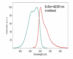
DiI, DiO, DiD and DiR dyes are a family of lipophilic fluorescent stains for labeling membranes and other hydrophobic structures. The fluorescence of these environment sensitive dyes is greatly enhanced when incorporated into membranes or bound to lipophilic biomolecules such as proteins although they are weakly fluorescent in water. Once applied to cells, these dyes diffuse laterally within the cellular plasma membranes, resulting in even staining of the entire cell at their optimal concentrations. DiOC6(3) is a green fluorescent membrane dye that has been used to detect mitochondrial membrane potential in live cells. The probe has also been employed in the analysis of apoptotic pathways by multiparametric flow cytometry. The dye has also been employed with the microfluidic Agilent 2100 bioanalyzer for assessing mitochondrial membrane potential. Wavelength Maxima: Excitation 482nm, Emission 504nm
Product Details
| Alternative Name: | 3,3’-Dihexyloxacarbocyanine iodide |
| |
| MW: | 572.5 |
| |
| CAS: | 53213-82-4 |
| |
| Purity: | ≥95% (HPLC) |
| |
| Solubility: | Soluble in DMSO. |
| |
| Shipping: | Ambient Temperature |
| |
| Long Term Storage: | -20°C |
| |
| Use/Stability: | Stable for at least one year after receipt when stored as recommended. |
| |
| Handling: | Protect from light. Keep cool and dry. |
| |
| Protocol: |
Prepare DMSO or EtOH stock solutions: The stock solutions should be prepared in DMSO or EtOH at 1-5mM. The unused portion of the stock solution should be stored at -20 °C. Avoid repeated freeze/thaw cycles.
Prepare working solutions: Dilute the stock solutions (from Step 1) into a suitable buffer such as serum-free culture medium, HBSS or PBS to make 1 to 5 µM working solutions. The final concentration of the working solution should be empirically determined for different cell types and/or experimental conditions. It is recommended to test at the concentrations that are at least over a tenfold range.
DiO and can be used with standard FITC.
Stain the cells in suspension:
-
Suspend cells at a density of 1 x 10⁶/ml in dye working solution (from Step 1.2).
-
Incubate at 37 °C for 2–20 minutes. The optimal incubation time varies depending on the cell type. Start by incubating for 20 minutes and subsequently optimize as necessary to obtain uniform labeling.
-
Centrifuge the labeled suspension tubes at 1000 to 1500 rpm for 5 minutes.
-
Remove the supernatant and gently resuspend the cells in pre-warmed (37 °C) growth medium.
-
Wash two more times as Steps 3 and 4.
Stain adherent cells:
-
Grow adherent cells on sterile glass coverslips.
-
Remove coverslips from growth medium and gently drain off excess medium. Place coverslip in a humidity chamber.
-
Pipet 100 μL of the dye working solution (from Step 1.2) onto the corner of a coverslip and gently agitate until all cells are covered.
-
Incubate the coverslip at 37 °C for 2–20 minutes. The optimal incubation time varies depending on the cell type. Start by incubating for 20 minutes and subsequently optimize as necessary to obtain uniform labeling.
-
Drain off the dye working solution and wash the coverslips two to three times with growth medium. For each wash cycle, cover the cells with pre-warmed growth medium, incubate for 5-10 minutes and then drain off the medium.
This product is a member of the CELLESTIAL® product line, reagents and assay kits comprising fluorescent molecular probes that have been extensively benchmarked for live cell analysis applications. CELLESTIAL® reagents and kits are optimal for use in demanding imaging applications, such as confocal microscopy, flow cytometry and HCS, where consistency and reproducibility are required. |
| |
| Regulatory Status: | RUO - Research Use Only |
| |

Cells labeled with DiO, can be analyzed using the conventional FL1 flow cytometer detection channels.
Please mouse over
Product Literature References
Revealing the suppressive role of protein kinase C delta and p38 mitogen-activated protein kinase (MAPK)/NF-κB axis associates with lenvatinib-inhibited progression in hepatocellular carcinoma in vitro and in vivo: C.H. Wu, et al; Biomed. Pharmacother.
145, 112437 (2022),
Abstract;
Amentoflavone Induces Apoptosis and Inhibits NF-ĸB-modulated Anti-apoptotic Signaling in Glioblastoma Cells: T.H. Yen, et al.; In Vivo
32, 279 (2018),
Abstract;
Full Text
Flagellin increases death receptor-mediated cell death in a RIP1-dependent manner: D. Hancz, et al.; Immunol. Lett.
193, 42 (2018),
Abstract;
Hyperforin Inhibits Cell Growth by Inducing Intrinsic and Extrinsic Apoptotic Pathways in Hepatocellular Carcinoma Cells: I.T. Chiang, et al.; Anticancer Res.
37, 161 (2017),
Abstract;
Analysis of apoptotic pathways by multiparametric flow cytometry: application to HIV infection: H. Lecoeur, et al.; Methods Enzymol.
442, 51 (2008),
Abstract;
Analysis of mitochondrial membrane potential in the cells by microchip flow cytometry: M. Kataoka, et al.; Electrophoresis
26, 3025 (2005),
Abstract;


