- Allows dual labeling with a variety of CELLESTIAL® fluorescent probes
- Minimal transfer of fluorescence from dye-labeled to unlabeled cells
- Suitable for long-term cell viability, cytotoxicity, cell adhesion, cell migration and cell-cell fusion assays
The CYTO-ID® Red Long-Term Cell Tracer Kit uses proprietary noncovalent cell labeling technology to stably incorporate a red fluorescent dye containing hydrophobic aliphatic chains into the cell membrane’s lipid bilayer. The dye may be loaded into cells by following the included protocol. The labeling buffer is isotonic for mammalian cells and contains no detergents or organic solvents. The appearance of labeled cells may vary depending upon the cell type from uniformly bright to punctuate. This difference is thought to relate to the extent of membrane internalization occurring after cell labeling. The CYTO-ID® Red Tracer Dye fluorescence is independent of pH within normally encountered physiologic ranges and fluorescence intensity per cell is typically unaffected by the ultimate pattern of dye distribution. The CYTO-ID® Red Tracer Dye is not toxic to cells, as determined using the benchmark MTT cell viability assay. The dye is well retained by cells for up to 96 hours after loading, and is passed to daughter cells upon mitosis. Since the dye does not covalently modify proteins within the cells, normal physiological responses are better preserved than with molecular probes based upon thiol-reactive chloromethyl-based or amine-reactive succinimidyl ester-based fluorescent dyes. Dual labeling is also possible using a variety of available CELLESTIAL® dyes. Labeled cells can be visualized by epifluorescence or confocal fluorescence microscopy. Additionally, dye-labeled and unlabeled cell populations can be analyzed by flow cytometry. No transfer of fluorescence to adjacent cells was observed after a prolonged 96-hour incubation period. This is in stark contrast to Calcein AM and BCECF AM, which are only retained within viable cells for a few hours at physiological temperatures. The kit is suitable for a variety of applications including long term cell viability, cytotoxicity, cell adhesion, cell migration and cell-cell fusion studies.
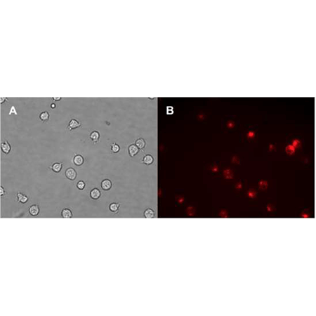
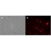
Composite bright-field (panel A) and fluorescence microscopy (panel B) images demonstrating staining of Jurkat cells with CYTO-ID Red Tracer Dye. Standard Texas Red filter set was used to image the membrane-bound signal.
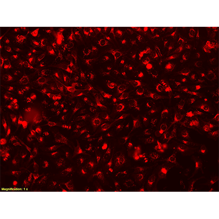
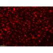
Fluorescence microscopy images demonstrating staining HeLa cells stained overnight with CYTO-ID® Red Tracer Dye. Standard Texas Red filter set was used to image the membrane-bound signal. (20x)
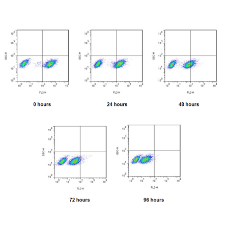
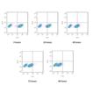
Flow Cytometry analysis of fluorescence of mixed population of Jurkat cells over time. Jurkat cells stained with CYTO-ID® Red Tracer dye were mixed with an unstained population of Jurkat cells and incubated over a 96 hour period.
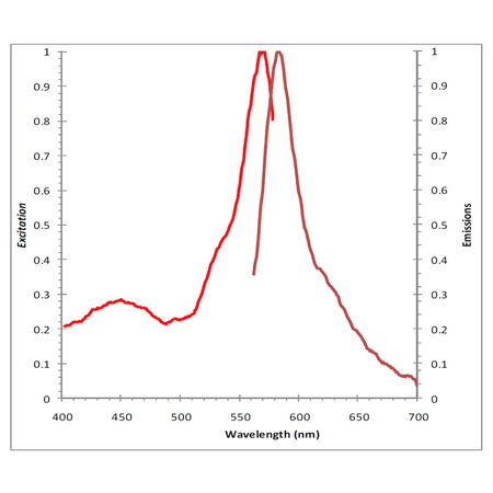
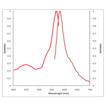
Fluorescence excitation (Ex 450nm, 570nm) and emission (Em 583nm) spectra for the CYTO-ID® Red Tracer Dye.
Please mouse over
Product Details
| Applications: | Flow Cytometry, Fluorescence microscopy, Fluorescent detection
|
| |
| Application Notes: | CYTO-ID® Red long-term cell tracer kit is suitable for a variety of applications including long term cell viability, cytotoxicity, cell adhesion, cell migration and cell-cell fusion studies. |
| |
| Quality Control: | A sample from each lot of CYTO-ID® Red long-term cell tracer kit is used to stain Jurkat cells and analyzed by flow cytometry using the procedures described in the user manual. Mean fluorescence of stained to unstained cells is greater than 5. |
| |
| Quantity: | 25 assays |
| |
| Use/Stability: | Store the kit at -20˚C in a non-frost free freezer, protected from light. Avoid multiple freeze-thaw cycles. With proper storage, the kit components are stable for one year from date of receipt. |
| |
| Handling: | Protect from light. Avoid freeze/thaw cycles. |
| |
| Shipping: | Dry Ice |
| |
| Short Term Storage: | -20°C |
| |
| Long Term Storage: | -20°C |
| |
| Contents: | CYTO-ID® Red Tracer Dye, 50 μl
4X Labeling Buffer, 12.5 ml
10X HBSS, 25 ml |
| |
| Technical Info/Product Notes: | The CYTO-ID® Red long-term cell tracer kit is a member of the CELLESTIAL® product line, reagents and assay kits comprising fluorescent molecular probes that have been extensively benchmarked for live cell analysis applications. CELLESTIAL® reagents and kits are optimal for use in demanding cell analysis applications involving confocal microscopy, flow cytometry, microplate readers and HCS/HTS, where consistency and reproducibility are required.
This product is manufactured and sold by ENZO LIFE SCIENCES, INC. for research use only by the end-user in the research market and is not intended for diagnostic or therapeutic use. Purchase does not include any right or license to use, develop or otherwise exploit this product commercially. Any commercial use, development or exploitation of this product or development using this product without the express prior written authorization of ENZO LIFE SCIENCES, INC. is strictly prohibited. |
| |
| Regulatory Status: | RUO - Research Use Only |
| |
Product Literature References
INBRX-120, a CD8α-targeted detuned IL-2 that selectively expands and activates tumoricidal effector cells for safe and durable in vivo responses: F. J. Sulzmaier, et al.; J. Immunother. Cancer
11, e006116 (2023),
Abstract;
Epigenetic modulation of immune synaptic-cytoskeletal networks potentiates γδ T cell-mediated cytotoxicity in lung cancer: R.R. Weng, et al.; Nat. Commun.
12, 2163 (2021),
Abstract;
Characterization and Cellular Internalization of Spherical Cellulose Nanocrystals (CNC) into Normal and Cancerous Fibroblasts: N.A.H. Shazali, et al.; Materials (Basel)
12, 3251 (2019),
Application(s): C6 and NIH3T3 cell tracing,
Abstract;
Time resolved 3D live-cell imaging on implants: A. Ingendoh-Tsakmakidis, et al.; PLoS One
13, e0205411 (2018),
Abstract;
Full Text
Nerve guidance conduit with a hybrid structure of a PLGA microfibrousbundle wrapped in a micro/nanostructured membrane: S.W. Peng, et al.; Int. J. Nanomedicine
12, 421 (2017),
Application(s):Tracking in vitro cells migration (immortalized neuron progenitor KT 98),
Abstract;
Full Text
Thioreductase-containing epitopes inhibit the development of type 1 diabetes in the NOD mouse model: E. Malek Abrahimians, et al.; Front. Immunol.
7, 67 (2016),
Application(s): Flow cytometry analysis using isolated mouse spleen B cells,
Abstract;
Full Text
Syngeneic murine ovarian cancer model reveals that ascites enriches for ovarian cancer stem-like cells expressing membrane GRP78: L. Mo, et al.; Mol. Cancer Ther.
14, 747 (2015),
Abstract;
An in vitro blood-brain barrier model combining shear stress and endothelial cell/astrocyte co-culture: Y. Takeshita, et al.; J. Neurosci. Methods
232, 165 (2014),
Application(s): Confocal microscopy using adult human astrocyte cells,
Abstract;
Full Text
Vascular endothelial growth factor-C modulates proliferation and chemoresistance in acute myeloid leukemic cells through an endothelin-1-dependent induction of cyclooxygenase-2: K.T. Hua, et al.; Biochim. Biophys. Acta
1843, 387 (2014),
Abstract;
Expanding the Ig superfamily code for laminar specificity in retina: expression and role of contactins: M. Yamagata, et al.; J. Neurosci.
32, 14402 (2012),
Abstract;
Full Text
General Literature References
Cell tracking 2007: A proliferation of probes and applications: P.K. Wallace & K.A. Muirhead; Immunol. Invest.
36, 527 (2007),
Abstract;
In: Living Color, Flow Cytometry and Cell Sorting Protocols: R.Y. Poon, et al.; R.A. Diamond & S. DeMaggio (eds.), Springer Verlag, New York NY 302 (2000), Book,
Fluorescent cell labeling for in vivo and in vitro cell tracking: P.K. Horan, et al.; Methods Cell Biol.
33, 469 (1990),
Abstract;
Stable cell membrane labeling: P.K. Horan & S.E. Slezak; Nature
340, 167 (1989),
Abstract;
Related Products


