The LYSO-ID® Green detection kit contains an acidic organelle-selective dye suitable for live-cell staining. Conventional fluorescent stains for acidic organelles, such as Acridine Orange (Prod. No. ENZ-52405), form metachromatic artifacts that interfere with multicolor imaging applications. LYSO-ID® Green dye generates emission profiles that can be multiplexed with other fluorophores. The dye accumulates in acidic compartments, such as endosomes, lysosomes, and secretory vesicles. Low micromolar concentrations of LYSO-ID® Green dye are sufficient for staining mammalian cells. This has been validated with the human cervical carcinoma cell line, HeLa, the human T-lymphocyte cell line, Jurkat, and the human bone osteosarcoma epithelial cell line, U2OS. The LYSO-ID® Green dye is a new green-emitting, cell-permeable small organic probe molecule that spontaneously localizes to live cell acidic organelles. It can be readily used in combination with other common UV and visible light excitable fluorescent dyes and various fluorescent proteins in multicolor imaging and detection applications. The LYSO-ID® Green dye is suitable for both short-term and long-term tracking studies. It emits in the FITC region of the visible light spectrum, and is highly resistant to photobleaching, concentration quenching and photoconversion. The LYSO-ID® Green detection kit is specifically designed for use with RFP-expressing cell lines, as well as cells expressing blue, cyan or orange fluorescent proteins (BFPs, CFPs, OFPs). A lysosome perturbation agent, chloroquine, is provided as a positive control for monitoring changes in lysosome number and volume. A nuclear counterstain is also provided in the kit to highlight this organelle as well.

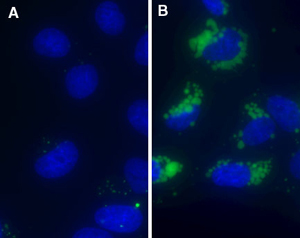
Figure 1: LYSO-ID® Green dye is a cell-permeant fluorescent probe that selectively associates with lysosomes and other acidic organelles (A). Cells pre-treated for 20 hours with weakly basic cell-permeant compounds, such as chloroquine, show dramatic increases in lysosome-like vesicle number and volume (B). Nuclei are counter-stained with Hoechst 33342 in the images.
Please mouse over
Product Details
| Applications: | Fluorescence microscopy, Fluorescent detection
|
| |
| Application Notes: | Specifically designed for use with RFP-expressing cell lines, as well as cells expressing blue, cyan or orange fluorescent proteins (BFPs, CFPs, OFPs). |
| |
| Quality Control: | A sample from each lot of LYSO-ID® Green detection kit is used to stain HeLa cells using the procedures described in the user manual. Analyzed by microscopy, the stained cells exhibit dramatic increase in lysosome-like vesicle number and volume in the chloroquine-treated HeLa cells. |
| |
| Use/Stability: | With proper storage, the kit components are stable for 1 year from the date of receipt. |
| |
| Handling: | Avoid freeze/thaw cycles. |
| |
| Shipping: | Blue Ice |
| |
| Short Term Storage: | -20°C |
| |
| Long Term Storage: | -80°C |
| |
| Contents: | LYSO-ID® Green detection reagent
Hoechst 33342 nuclear staining
Chloroquine control
10x Assay buffer |
| |
| Technical Info/Product Notes: | The LYSO-ID® Green detection kit is a member of the CELLESTIAL® product line, reagents and assay kits comprising fluorescent molecular probes that have been extensively benchmarked for live cell analysis applications. CELLESTIAL® reagents and kits are optimal for use in demanding cell analysis applications involving confocal microscopy, flow cytometry, microplate readers and HCS/HTS, where consistency and reproducibility are required. |
| |
| Regulatory Status: | RUO - Research Use Only |
| |
Product Literature References
A novel glucosylceramide synthase inhibitor attenuates alpha synuclein pathology and lysosomal dysfunction in preclinical models of synucleinopathy: M. Cosden, et al.; Neurobiol. Dis.
159, 105507 (2021),
Abstract;
Areca nut extract (ANE) inhibits the progression of hepatocellular carcinoma cells via activation of ROS production and activation of autophagy: P.L. Wei, et al.; Int. J. Med. Sci.
18, 3452 (2021),
Abstract;
Autophagy-dependent glutaminolysis drives superior IL21 production in HIV-1-specific CD4 T cells: H. Loucif, et al.; Autophagy
1, 18 (2021),
Abstract;
Biomimetic cell membrane-coated DNA nanoparticles for gene delivery to glioblastoma: S. Han, et al.; J. Control. Release
338, 22 (2021),
Abstract;
Lipophagy confers a key metabolic advantage that ensures protective CD8A T-cell responses against HIV-1: H. Loucif, et al.; Autophagy
17, 3408 (2021),
Abstract;
Full Text
Methyl gallate, gallic acid-derived compound, inhibit cell proliferation through increasing ROS production and apoptosis in hepatocellular carcinoma cells: C.Y. Huang, et al.; PLoS One
16, e0248521 (2021),
Abstract;
Full Text
RFP-based method for real-time tracking of invasive bacteria in a heterogeneous population of cells: R.O. Akinsola, et al.; Anal. Biochem.
634, 114432 (2021),
Abstract;
Lysosomal dysfunction and autophagy blockade contribute to MDMA-induced neurotoxicity in SH-SY5Y neuroblastoma cells: I.H. Li, et al.; Chem. Res. Toxicol.
33, 903 (2020),
Abstract;
Bromelain inhibits the ability of colorectal cancer cells to proliferate via activation of ROS production and autophagy: T.C. Chang, et al.; PLoS One
14, e0210274 (2019),
Application(s): Flow cytometry using HCT116 and HT-29 cells,
Abstract;
Full Text
Propyl gallate inhibits hepatocellular carcinoma cell growth through the induction of ROS and the activation of autophagy: P.L. Wei, et al.; PLoS One
14, e0210513 (2019),
Abstract;
Full Text
Targeting of cathepsin C induces autophagic dysregulation that directs ER stress mediated cellular cytotoxicity in colorectal cancer cells: T.P. Khaket, et al.; Cell. Signal.
46, 92 (2018),
Abstract;
Imipramine blue sensitively and selectively targets FLT3-ITD positive acute myeloid leukemia cells: J. Metts, et al.; Sci. Rep.
7, 4447 (2017),
Abstract;
Full Text
Autophagic lysosome reformation dysfunction in glucocerebrosidase deficient cells: relevance to Parkinson Disease: J. Magalhaes, et al.; Hum. Mol. Genet.
25, 3432 (2016),
Application(s): Fluorescence microscopy and microplate reader using neuroblastoma SH-SY5Y cells and mouse cortical neurons,
Abstract;
Ferritin-Mediated siRNA Delivery and Gene Silencing in Human Tumor and Primary Cells: L. Li, et al.; Biomaterials
98, 143 (2016),
Application(s): Lysosomal Detection,
Abstract;
A cancer cell-activatable aptamer-reporter system for one-step assay of circulating tumor cells: Z. Zeng, et al.; Mol. Ther. Nucleic Acids
3, e184 (2014),
Application(s): Confocal microscopy,
Abstract;
Full Text
Ambroxol improves lysosomal biochemistry in glucocerebrosidase mutation-linked Parkinson disease cells: A. McNeill, et al.; Brain
137, 1481 (2014),
Application(s): Detection using a microplate reader,
Abstract;
Full Text
Inhibition of beclin1 affects the chemotherapeutic sensitivity of osteosarcoma: W. Wu, et al.; Int. J. Clin. Exp. Pathol.
7, 7114 (2014),
Application(s): Fluorescence microscopy on human osteosarcoma cell line,
Abstract;
Full Text
Multifluorescence live analysis of herpes simplex virus type-1 replication: M. Seyffert, et al.; Methods Mol. Biol.
1144, 235 (2014),
Abstract;
A novel sulindac derivative inhibits lung adenocarcinoma cell growth through suppression of Akt/mTOR signaling and induction of autophagy: E. Gurpinar, et al.; Mol. Cancer Ther.
12, 663 (2013),
Abstract;
General Literature References
Photoconversion of Lysotracker Red to a green fluorescent molecule: E.C. Freundt, et al.; Cell Res.
17, 956 (2007),
Abstract;
Resolving vesicle fusion from lysis to monitor calcium-triggered lysosomal exocytosis in astrocytes: J.K. Jaiswal, et al.; PNAS
104, 14151 (2007),
Abstract;
Systematic colocalization errors between acridine orange and EGFP in astrocyte vesicular organelles: F. Nadrigny, et al.; Biophys. J.
93, 969 (2007),
Abstract;
Disruptive effect of chloroquine on lysosomes in cultured rat hepatocytes: A. Michihara, et al.; Biol. Pharm. Bull.
28, 947 (2005),
Abstract;
Photo-oxidative disruption of lysosomal membranes causes apoptosis of cultured human fibroblasts: U.T. Brunk, et al.; Free Radic. Biol. Med.
23, 616 (1997),
Abstract;
Related Products


