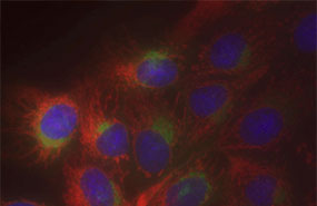
Figure: Detection of full-length human RIP3 (aa 1-519) with MAb to RIP3 (human) (Rippy-3) (Prod. No.
ALX-804-155) in a cell lysate control of mock transfected cells (HEK 293T) (Prod. No.
ALX-840-600) (lane 1) and HEK 293T cells transfected with a full-length human RIP3 expression plasmid (Prod. No.
ALX-840-608) (lane 2).
Method: 13µl of water and 2µl of DTT (100mM) were added to 5µl of cell lysate (Prod. No.
ALX-840-608) (corresponding to 2x10
4 cells) and boiled 5 min before being loaded on a 12% SDS-PAGE gel. The gel was run at 200V and proteins transferred to nitrocellulose (Amersham Biosciences, Prod. No. 25006565) for 1 hour at 100V in transfer buffer (3g/lt trizma-base, 14.4g/lt glycine, 20% methanol at pH 8.3). The nitrocellulose membrane was then blocked in PBS-Tween 0.1%, milk 4% at 4°C and incubated with MAb to RIP3 (human) (Rippy-3) (Prod. No.
ALX-804-155) at 1µg/ml in PBS-Tween 0.1%, milk 4% for 1 hour at RT. After three washes in PBS-Tween 0.1%, the membrane was incubated with goat-mouse horseradish peroxydase (Prod. No. ALX-211-203) diluted 1:5’000 in PBS-Tween 0.1%, milk 4% for 45 min. at RT. After three washes in PBS-Tween 0.1%, the membrane is revealed in ECL solution (Pierce, Prod. No. 34080). The membrane was exposed for 30 sec with a Fujimedical X-Ray film (Fujifilm, Prod. No. 57164053)."