Alternative Name: | MAPK2/3, MAP kinase 2/3 |
|
Clone: | G15-B |
|
Host: | Rabbit |
|
Isotype: | IgG |
|
Immunogen: | Synthetic peptide comprising the pThr-Glu-pTyr motif in activated ERK1 and ERK2. |
|
UniProt ID: | P27361 |
|
Species reactivity: | Human, Mouse, Rat
|
|
Specificity: | Recognizes the pThr-Glu-pTyr motif of activated ERK1 and ERK2. |
|
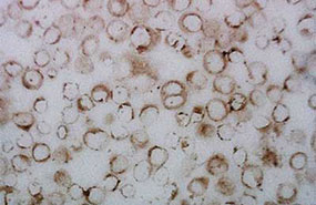
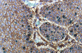
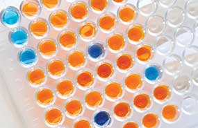
Applications: | ELISA, IHC (FS), IHC (PS), IP, WB
|
|
Recommended Dilutions/Conditions: | ELISA (1:20,000-1:100,000) Western Blot (1:5,000; Wash buffer: 1x TRIS buffered saline (TBS) containing 0.1% Triton X-100, Blocking buffer: 1x TBS containing 0.1% Triton X-100 and 5% BSA (used with the primary antibody). Incubate the membrane with antibody diluted in blocking buffer for 2 hours at room temperature) Suggested dilutions/conditions may not be available for all applications. Optimal conditions must be determined individually for each application. |
|
Purity Detail: | Major clone of IgGs obtained from immunoaffinity purified immunoglobulins corresponding to immunogenic peptide. |
|
Formulation: | Liquid. In 20mM TRIS/HCl, pH 8.0, containing 10mg/ml BSA and 0.05% sodium azide. |
|
Use/Stability: | Stable for at least 12 months after receipt when stored as recommended. |
|
Handling: | Avoid freeze/thaw cycles. |
|
Shipping: | Blue Ice Not Frozen |
|
Long Term Storage: | +4°C |
|
Technical Info/Product Notes: | Clonal Rabbit Antibody is a pure homogenous fraction of rabbit immunoglobulin (IgG) which corresponds to a unique linear epitope on the target protein. This epitope is designed after a detailed analysis of the antigen structure, regulatory post-translational modifications, protein/protein interaction domain and protein folding. The chosen sequence (peptide) is synthesized and the rabbit is immunized. The clone is then separated from the crude antiserum by "in vitro cloning technology". The resulting product is monospecific and characterized by superior specificity, affinity and avidity. |
|
Regulatory Status: | RUO - Research Use Only |
|
Figure: Western blot analysis of ERK1/2 activation in untreated PC12 cells (A), cells treated with EGF (100ng/ml, 5min) (B), PMA (100nM, 30min, in serum free DMEM) (C), and EGF (100ng/ml, 5min with 10% FBS in DMEM) (D). Wells were equally loaded with 50μg of whole cell lysate proteins/well. Western blot was performed and kindly provided by Dr. Martina Takáčová from Intitute of Virology, Slovak Academy of Sciences, Bratislava, Slovakia.