Replaces Prod. #: BML-SA264
This reagent (MAb F7-26) allows the important discrimination between apoptosis and necrosis. In this respect it can be regarded as a unique research tool superior to TUNEL staining. The specificity of the assay is based upon the high sensitivity of DNA to thermal denaturation in condensed chromatin of apoptotic cells. Although there is no change of the original product there have been important improvements in the staining protocol involving a formamide-based DNA-denaturation. This is now more convenient to use, yielding a sharper contrast between apoptotic cells and controls. The increased sensitivity of MAb F7-26 allows omission of a previously recommended Biotin/Streptavidin amplification step, a source of background problems. The APOSTAIN assay is now applicable for cold (4°C) formalin fixed tissue.
Product Details
Alternative Name: | Single-stranded DNA |
|
Clone: | F7-26 |
|
Host: | Mouse |
|
Isotype: | IgM |
|
Immunogen: | Calf thymus ssDNA. |
|
Species reactivity: | Species independent
|
|
Specificity: | Recognizes deoxycytidine of ssDNA of at least 25-30 bases in length in the absence of any reactivity to double-stranded DNA; not species-specific. Specifically detects apoptotic cells; necrotic cells are not recognized. |
|
Applications: | Flow Cytometry, ICC, IHC (FS), IHC (PS)
|
|
Recommended Dilutions/Conditions: | Immunohistochemistry (frozen sections, paraffin sections - important: applicable for cold fixation at +4°C - for reference see Manual) Suggested dilutions/conditions may not be available for all applications. Optimal conditions must be determined individually for each application. |
|
Quantity: | 200µl/20 tests; 1ml/100 tests |
|
Formulation: | Liquid. In PBS containing 5% fetal bovine serum (FBS). |
|
Shipping: | Dry Ice |
|
Long Term Storage: | -80°C |
|
Regulatory Status: | RUO - Research Use Only |
|
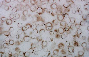
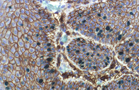
Figure: Photomicrographs of thymus from a mouse treated with hydrocortisone (left) and of a control treated animal (right). Sections of formalin-fixed tissues were heated in formamide, stained with
MAb F7-26 (Prod. No.
ALX-804-192) and counterstained with hematoxylin. Brown nuclei represent antibody stained apoptotic cells with condensed chromatin and fragmented nuclei."
Please mouse over
Product Literature References
Linoleic Acid Upregulates Microrna-494 to Induce Quiescence in Colorectal Cancer: R. Ogata, et al.; Int. J. Mol. Sci.
23, 225 (2022),
Abstract;
Evading immune surveillance via tyrosine phosphorylation of nuclear PCNA: Y.L. Wang, et al.; Cell Rep.
36, 109537 (2021),
Abstract;
The Role of Pseudomonas aeruginosa ExoY in an Acute Mouse Lung Infection Model: C. Kloth, et al.; Toxins (Basel)
10, 185 (2018),
Abstract;
Full Text
Cementocyte cell death occurs in rat cellular cementum during orthodontic tooth movement: H. Matsuzawa, et al.; Angle Orthod.
87, 416 (2017),
Abstract;
Full Text
Evidence of ubiquitous in vivo and in vitro expression of pro-apoptotic Smac/DIABLO protein in meningioma cell lines: C. Pfister, et al.; Oncol. Rep.
21, 1181 (2009),
Abstract;
Extracellular ATP induces retinal photoreceptor apoptosis through activation of purinoceptors in rodents: T. Puthussery, et al.; J. Comp. Neurol.
513, 430 (2009),
Abstract;
Cathepsin-B-dependent apoptosis triggered by antithymocyte globulins: a novel mechanism of T-cell depletion: M. C. Michallet, et al.; Blood
102, 3719 (2003),
Abstract;
Caspase-independent phosphatidylserine exposure during apoptosis of primary T lymphocytes: C. Ferraro-Peyret, et al.; J. Immunol.
169, 4805 (2002),
Abstract;
Full Text
Effects of dexamethasone on apoptosis-related cell death after spinal cord injury: M. Zurita, et al.; J. Neurosurg.
96, 83 (2002),
Abstract;
Imbalance between apostain expression and proliferative index can predict survival in primary glioblastoma: J. Vaquero, et al.; Acta Neurochir. (Wien)
144, 151 (2002),
Abstract;
Apoptosis: one of the mechanisms that maintains unresponsiveness of the intestinal mucosal immune system: P. Bu, et al.; J. Immunol.
166, 6399 (2001),
Abstract;
Identification of apoptotic cells by formamide-induced dna denaturation in condensed chromatin: O.S. Frankfurt & A. Krishan; J. Histochem. Cytochem.
49, 369 (2001),
Abstract;
Prediction of imminent complications in HIV-1-infected patients by markers of lymphocyte apoptosis: J.C. Wasmuth, et al.; J. Acquir. Immune Defic. Syndr.
23, 44 (2000),
Abstract;
DNA strand breaks in Alzheimer's disease: E. Adamec, et al.; Brain Res.
849, 67 (1999),
Abstract;
In vivo amifostine (WR-2721) prevents chemotherapy-induced apoptosis of peripheral blood lymphocytes from cancer patients: M. Provinciali, et al.; Life Sci.
64, 1525 (1999),
Abstract;
Detection of apoptotic cells with monoclonal antibodies to single-stranded DNA: O.S. Frankfurt; Apoptosis Detection and Assay Methods (Eds L. Zhu & J. Chun) 47 (1998),
Early T-cell apoptosis and Fas expression during antiretroviral therapy in individuals infected with human immunodeficiency virus-1: S.P. Aries, et al.; Scand. J. Immunol.
48, 86 (1998),
Abstract;
Apoptosis in breast carcinomas detected with monoclonal antibody to single-stranded DNA: relation to bcl-2 expression, hormone receptors, and lymph node metastases: O.S. Frankfurt et al.; Clin. Cancer Res.
3, 465 (1997),
Abstract;
Monoclonal antibody to single-stranded DNA is a specific and sensitive cellular marker of apoptosis: O.S. Frankfurt, et al.; Exp. Cell. Res.
226, 387 (1996),
Abstract;
A cautionary note on the use of the TUNEL stain to determine apoptosis: C. Charriaut-Marlangue & J. Ben-Ari; Neuroreport
7, 61 (1995),
Abstract;
In Situ detection of fragmented DNA (TUNEL assay) fails to discriminate among apoptosis, necrosis and autolytic cell death: A cautionary note: B. Grasl-Kraupp, et al.; Hepatology
21, 1465 (1995),
Abstract;
Detection of apoptosis in leukemic and breast cancer cells with monoclonal antibody to single-stranded DNA: O.S. Frankfurt; Anticancer Res.
14, 1861 (1994),
Abstract;
Decreased stability of DNA in cells treated with alkylating agents: O.S. Frankfurt; Exp. Cell. Res.
191, 181 (1990),
Abstract;
Detection of DNA damage in individual cells by flow cytometric analysis using anti-DNA monoclonal antibody: O.S. Frankfurt; Exp. Cell. Res.
170, 369 (1987),
Abstract;