-
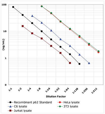
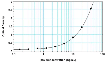
Highly sensitive assay, measure as little as 100 pg/ml of p62
-
Fully quantitative results surpass semi-quantitative Western blot analysis
-
Higher throughput format, assay up to 40 samples in duplicate in just 3 hours
-
Easy-to-use liquid color-coded reagents reduce errors
The p62 ELISA kit is a colorimetric, immunometric immunoassay kit with results in 3 hours.
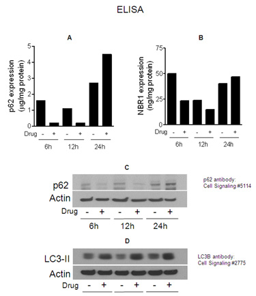
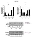
Correlation of p62 (Prod. No. ADI-900-212) and NBR1 (Prod. No. ADI-900-211) immunoassays to autophagy induction. MDA-MB-231 human breast cancer cells were treated with 2μM of withaferin A (WA), an autophagy inducing drug. Cells were harvested at 6, 12 and 24 hours post-treatment and lysed in RIPA cell lysis buffer 2 containing protease inhibitors and DNase. Cell lysates were clarified by centrifugation and analyzed in p62 assay (Figure 3A) and NBR1 assay (Figure 3B). Concentration of antigen was normalized to total cellular protein. Cell lysates were resolved by SDS-PAGE and analyzed by Western blot using anti-p62 antibody from Cell Signaling, CN #5114 (Figure 3C) or anti-LC3 antibody from Cell Signaling, CN #2775 (Figure 3D). Signal intensity in both Western blots were compared to actin levels and numbers above band represent changes in protein levels relative to corresponding DMSO-treated control.
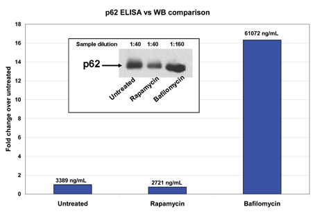
Stimulation Assay, p62 ELISA vs WB comparison: Human HeLa cells were treated for 24 hours with 800nM Rapamycin, an inducer of autophagy, or 800nM Bafilomycin A1, an inhibitor of vacuolar ATPase which leads to an accumulation of autophagosomal structures. Cells were then harvested, washed and lysed at 5x10⁶ per mL RIPA Cell Lysis Buffer 2 following the procedure in the Cell Lysate Preparation section. These samples were diluted and resolved on an 80-12% Tris-glycine gel, transferred to nitrocellulose membrane and probed for p62. The same cell lysates were also diluted in assay buffer and run in this kit. The Western Blot provided a visual for both the decrease and increase in p62 levels after treatment of cells in culture. The p62 ELISA kit was able to assign values to the amounts of p62 protein (numbers above each bar) thus allowing a quantitative comparison of the decrease with Rapamycin treatment (~25%) and the increase with Bafilomycin A1 treatment (~1600%).
Please mouse over
Product Details
| Alternative Name: | Sequestosome 1 |
| |
| Sensitivity: | 100pg/ml (range 625 - 40,000pg/ml) |
| |
| Assay Time: | 3 hours |
| |
| Applications: | ELISA, Colorimetric detection
|
| |
| Application Notes: | For the quantitative determination of human, rat and mouse p62 in cell lysate samples. Cited sample type includes in PBMC lysates. |
| |
| Wavelength: | 450 nm |
| |
| Species reactivity: | Human, Mouse, Rat
|
| |
| Crossreactivity: | No cross reactivity. |
| |
| Use/Stability: | Store all components at +4ºC, except Standard at -80ºC. |
| |
| Shipping: | Dry Ice |
| |
| Contents: | Microtiter Plate, Antibody, Conjugate, Assay Buffer 13, Wash Buffer Concentrate, Standard, TMB Substrate, Stop Solution 2, RIPA Cell Lysis Buffer 2 |
| |
| Scientific Background: | The generic term ‘‘autophagy’’ comprises several processes by which the lysosome acquires cytosolic cargo, with three types of autophagy being discerned in the literature: (1) macroautophagy, characterized by the formation of a crescent-shaped structure (the phagophore) that expands to form the double-membrane autophagosome, capable of fusion with the lysosome; (2) microautophagy, in which lysosomes invaginate and directly sequester cytosolic components; and (3) chaperone-mediated autophagy (CMA), which involves translocation of unfolded proteins across the lysosomal membrane.
Upregulation of autophagy pathways occurs in response to extra- or intracellular stress and signals such as starvation, growth factor deprivation, ER stress and pathogen infection. Malfunction of these pathways is linked to various human pathologies including cancer, neurodegeneration and infectious diseases.
Selective macroautophagy describes the pathway of self-degradation of whole cellular components, protein aggregates or unusually long-lived proteins; in which double-membrane autophagosomes sequester organelles, ubiquitinylated proteins or ubiquitinylated protein aggregates and subsequently fuse with lysosomes for breakdown by resident hydrolases. Autophagic clearance of protein aggregates requires the ubiquitin-binding receptors p62 and NBR1.
The p62 protein, also known as sequestosome 1 (SQSTM1), has a dual functionality as both a scaffold protein and aiding in trafficking for protein degradation. It can polymerize and bind to NBR-1 via a PB1 (Phox and Bem1) domain, interact with ubiquitinated proteins linking them to the autophagic machinery via a UBA (ubiquitin-associated) domain and bind to the LC3II protein of the autophagy pathway through an LIR (LC3 interacting region) motif. p62 provides a key link between the ubiquitin-proteasome system (UPS) and autophagy by facilitating autophagic degradation of ubiqutinated proteins, decreasing aggregation of misfolded and non-functional proteins within cells, resulting in enhanced cellular survival characteristics. Because p62 accumulates when autophagy is inhibited, and decreased levels can be observed when autophagy is induced, p62 may be used as a biomarker to study autophagic flux. Also, p62 has been implemented in neurodegenerative diseases such as Parkinson's and Alzheimer's and in the skeletal disorder Paget’s disease of bone, establishing the p62 protein as a potential therapeutic target. |
| |
| UniProt ID: | Q13501 (human) |
| |
| Regulatory Status: | RUO - Research Use Only |
| |
| Compatibility: | This product is compatible with the Absorbance 96 Plate Reader.
 |
| |
Product Literature References
Adaptive autophagy reprogramming in Schwann cells during peripheral demyelination: Y.R. Jo, et al.; Cell. Mol. Life Sci.
80, 34 (2023),
Abstract;
Alterations in Proteostasis System Components in Peripheral Blood Mononuclear Cells in Parkinson Disease: Focusing on the HSP70 and p62 Levels: J.D. Vavilova, et al.; Biomolecules
12, 493 (2022),
Abstract;
AMPK/mTOR-driven autophagy & Nrf2/HO-1 cascade modulation by amentoflavone ameliorates indomethacin-induced gastric ulcer: M.F. Balaha, et al.; Biomed. Pharmacother.
151, 113200 (2022),
Abstract;
Increase of autophagy marker p62 in the placenta from pregnant women with preeclampsia: V.R. Ribeiro, et al.; Hum. Immunol.
S0198, 8859 (2022),
Abstract;
Alterations of the 70 kDa heat shock protein (HSP70) and sequestosome-1 (p62) in women with breast cancer: T. Orfanelli, et al.; Sci. Rep.
11, 22220 (2021),
Abstract;
Cellular Toxicity Mechanisms and the Role of Autophagy in Pt(IV) Prodrug-Loaded Ultrasmall Iron Oxide Nanoparticles Used for Enhanced Drug Delivery: L.G. Romero, et al.; Pharmaceutics
13, 1730 (2021),
Abstract;
An increase in p62/NBR1 levels in melioidosis patients of Sri Lanka exhibit a characteristic of potential host biomarker: K. U. Saikh, et al.; J. Med. Microbiol.
69, 1240 (2020),
Abstract;
Full Text
Modulation of Autophagy Influences the Function and Survival of Human Pancreatic Beta Cells Under Endoplasmic Reticulum Stress Conditions and in Type 2 Diabetes: M. Bugliani, et al.; Front. Endocrinol. (Lausanne)
10, 52 (2019),
Application(s): Human primary islet cell lysates,
Abstract;
Full Text
Autophagy in human skin fibroblasts: impact of age: H.S. Kim, et al.; Int. J. Mol. Sci.
19, 2254 (2018),
Application(s): ELISA using cell lysates and culture supernatants,
Abstract;
The first-in-class alkylating deacetylase inhibitor molecule tinostamustine shows antitumor effects and is synergistic with radiotherapy in preclinical models of glioblastoma: C. Festuccia, et al.; J. Hematol. Oncol.
11, 32 (2018),
Abstract;
Full Text
Autophagy Biomarkers Beclin 1 and p62 are Increased in Cerebrospinal Fluid after Traumatic Brain Injury: A. K. Au, et al.; Neurocrit. Care
26, 348 (2017),
Application(s): ELISA using human cerebrospinal fluid (CSF) samples,
Abstract;
Macrophage Migration Inhibitory Factor (MIF) Deficiency Exacerbates Aging-Induced Cardiac Remodeling and Dysfunction Despite Improved Inflammation: Role of Autophagy Regulation: X. Xu, et al.; Sci. Rep.
6, 22488 (2016),
Application(s): Western blot,
Abstract;
Full Text
Activity of BKM120 and BEZ235 against Lymphoma Cells: M. Civallero, et al.; Biomed Res. Int.
2015, Article ID 870918 (2015),
Application(s): Autophagy Detection in cell lysates,
Abstract;
Full Text
STAT3 down regulates LC3 to inhibit autophagy and pancreatic cancer cell growth: J. Gong, et al.; Oncotarget
5, 2529 (2014),
Application(s): Levels of p62 in human pancreatic cancer cells Capan-2,
Abstract;
Full Text
Impaired autophagy by soluble endoglin, under physiological hypoxia in early pregnant period, is involved in poor placentation in preeclampsia: A. Nakashima, et al.; Autophagy
9, 303 (2013),
Abstract;
Inhibition of autophagy by sera from pregnant women: T.T. Kanninen, et al.; Reprod. Sci.
20, 1327 (2013),
Application(s): EIA using PBMC lysates,
Abstract;
General Literature References
A role for NBR1 in autophagosomal degradation of ubiquitinated substrates: V. Kirkin, et al.; Mol. Cell
33, 505 (2009),
Abstract;
A role for ubiquitin in selective autophagy: V. Kirkin, et al.; Mol. Cell
34, 259 (2009),
Abstract;
Monitoring autophagic degradation of p62/SQSTM1: G. Bjorkoy, et al.; Methods Enzymol.
452, 181 (2009),
Abstract;
NBR1 cooperates with p62 in selective autophagy of ubiquitinated targets: V. Kirkin, et al.; Autophagy
5, 732 (2009),
Abstract;
Autophagy fights disease through cellular self-digestion: N. Mizushima, et al.; Nature
451, 1069 (2008),
Abstract;
p62/SQSTM1 binds directly to Atg8/LC3 to facilitate degradation of ubiquitinated protein aggregates by autophagy: S. Pankiv, et al.; J. Biol. Chem.
282, 24131 (2007),
Abstract;
Signaling, polyubiquitination, trafficking, and inclusions: sequestosome 1/p62's role in neurodegenerative disease: M.W. Wooten, et al.; J. Biomed. Biotechnol.
2006, 62079 (2006),
Abstract;
Evidence for p62 aggregate formation: role in cell survival: M.G. Paine, et al.; FEBS Lett.
579, 5029 (2005),
Abstract;
p62/SQSTM1 forms protein aggregates degraded by autophagy and has a protective effect on huntingtin-induced cell death: G. Bjorkoy, et al.; J. Cell Biol.
171, 603 (2005),
Abstract;
Structure of the ubiquitin-associated domain of p62 (SQSTM1) and implications for mutations that cause Paget's disease of bone: B. Ciani, et al.; J. Biol. Chem.
278, 37409 (2003),
Abstract;
Early accumulation of p62 in neurofibrillary tangles in Alzheimer's disease: possible role in tangle formation: E. Kuusisto, et al.; Neuropathol. Appl. Neurobiol.
28, 228 (2002),
Abstract;
Related Products


