In the last decade, there have been numerous breakthroughs in our ability to understand the potential causes and effects of cancerous diseases as well as the immune responses to internal and external stimuli. Cancer is defined as a complex and heterogeneous group of diseases characterized by uncontrolled and disorderly proliferation of cells. Biomarkers in cancer can provide the ability to measure features that characterize normal or abnormal biological states in our bodies. Whether its analyzing biomolecules such as DNA, RNA, protein, and peptide, every cell type has unique molecular signatures and identifiable characteristics such as levels or activities. Combining this characteristic of tumor cells with the ability to specifically assay for these cancer biomolecules allows for powerful methods of detection.
Enzyme-linked Immunosorbent Assay (ELISA) and Immunohistochemistry (IHC) are two ways in which we are able to quantify the effects of the surround tissues or cells in response to the tumor. ELISA provides a biochemical approach which measures the concentration of a specific analytes through antibody analyte binding affinity and colorimetric development. While IHC involves the process of identifying antigen of interest by using the principle of antibodies binding specifically to antigen in biological tissues. Both of these applications are widely used in cancer research as they augment our ability to identify abnormalities.
Applications with Immunohistochemistry (IHC)
KI-67
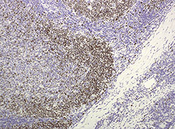
Ki-67 is a nuclear non-histone protein present in all active phases of the cell cycle, except the G0 phase. Ki-67 is present in all proliferating cells, and its role as a proliferation marker has attracted considerable interest. In interphase cells, Ki-67 primarily localizes to the nucleolus, whereas during mitosis, it coats the chromosomes. Specifically, Ki-67 is required for formation of the mitotic perichromosomal layer that coats mitotic chromosomes. In this layer, Ki-67's large size and highly positively charged amino acid composition keep individual mitotic chromosomes dispersed rather than aggregated upon nuclear envelope disassembly, thereby ensuring normal kinetics of anaphase progression. Although the antigen has also been associated with ribosomal RNA transcription, it is strongly linked to cell proliferation and has thus been indicated as an effective marker in grading the proliferation rate of tumors, including those of the brain, breast, cervix, and prostate. By immunostaining with the monoclonal antibody Ki-67, it is possible to assess the growth fraction of neoplastic cell populations. However, to date no standard operating procedure or generally accepted cut-off definition for Ki-67 exists. For this reason, both the inter-laboratory and the inter-study comparability of Ki-67 are limited. Therefore, Ki-67 is not implemented in standard routine pathology so far.
LAG-3
Lymphocyte-activation gene 3 (LAG-3 or CD233) is complex transmembrane protein that expressed on activated T Cells. LAG-3 is an immunoglobulin superfamily. It is a structural homolog to CD4 that binds to major histocompatibility complex (MHC) class II molecules on APCs. Although the signaling mechanism of LAG-3 is still mostly unknown, LAG-3 has also shown that it negatively regulates cellular proliferation, activation, and homeostasis of T cells, and it also helps maintain CD8+ T cells in a tolerogenic state and, working with PD-1, helps maintain CD8 exhaustion during chronic viral infection. LAG3 is considered to be involved in the maturation and activation of dendritic cells, and it also inhibits CD8+ effector T cell function independently of its role on Treg cells. For its ligand, MHC class II molecules, which are upregulated on some epithelial cancers, LAG-3 is immuno-inhibitory much like CTLA-4 and PD-1 but promotes a less potent immunosuppressive effect on T-cells.
Her2/Neu
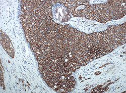
Human epidermal growth factor receptor 2 (HER2/Neu) is one of the four members that comprise the ErbB family of receptor tyrosine kinases. It is typically associated with cell proliferation, survival, and opposes apoptosis. Ligand binding to extracellular domains causes HER2 to undergo dimerization and transphosphorylation of intracellular domain tyrosine residues. Subsequent phosphorylation of dock intracellular molecules results in downstream activation of other signaling pathways such as MAPK, PI3K/Akt, and PKC. Relating to cancer, HER2 is a major proto-oncogene that is overexpressed in up to 20-30% of breast cancers in women. Evidence suggests that HER2 amplification occurs early in human breast tumorigenesis and can have up to 40-100 fold increase in HER2 protein expression on tumor cell surfaces. Diagnosis of HER2-positive breast cancer amplification can be reliably done with IHC testing. Results will either be 0 (negative), 1+ (negative), 2+ (borderline), or 3+ (positive). Extrapolating these findings helps determine prognosis and establish suitability for treatments such as Trastuzumab, a murine IgG mAb that binds to the HER2 extracellular domains.
Immunoassay for Cancer Research
p53

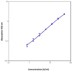
Figure 3: Standard curve of the p53 (human) ELISA kit (ALX-850-057) depicting the detection range (0.33U/ml – 50U/ml).
|
p53 is the most commonly mutated tumor suppressor gene for human cancer research, by de-regulating the p53 to operate as a transcription factor that activates genes regulating cell cycle checkpoints to induce cell cycle arrest or apoptosis in response to DNA damage. P53 is a phosphoprotein made up of 393 amino acid. P53 is considered a stress response gene and acts to induce cell cycle arrest or apoptosis in response to DNA damage. p53 as a transcription factor induces the expression of p21WAF1/CIP1/Sdi1 leading to G1 arrest of the cell. p53 is known to be regulated by phosphorylation by a number of specific protein kinases. Activation by autoproteolysis has been shown. The role in which p53 plays therefore, quantifying the amount of p53 biomarker (or lack thereof) is an attractive target of interest when studying cancer to examine the expression relative to the progression of transformed cells. Enzo Life Sciences offers our
p53 (human) ELISA kit, which is a colorimetric immuno-metric assay capable of quantifying p53 in cell culture supernatants, human serum, plasma, and other biological fluids.
VEGF
Angiogenesis, or new vessel formation, is an essential physiological process for embryologic development, normal growth, and tissue repair. Angiogenesis is tightly regulated at the molecular level; however, this process is dysregulated in several pathological conditions such as cancer. Vascular endothelial growth factor (VEGF) is a homodimeric glycoprotein mitogen that plays a crucial role in angiogenesis. The VEGF family consists of five distinct ligands, VEGF-A, -B, -C and -D and placental growth factor, or PlGF. Synthesis of VEGF is up-regulated by a variety of growth factors, cytokines and hypoxia. The production of VEGF and other growth factors by the tumor results in the 'angiogenic switch', where new vasculature is formed in and around the tumor, allowing it to grow exponentially. Tumor vasculature formed under the influence of VEGF is structurally and functionally abnormal. The discovery of VEGF as a major driver of tumor
angiogenesis has led to develop novel therapeutics aimed at inhibiting its activity. The first anti-angiogenic cancer therapeutic, the neutralizing antibody bevacizumab, targets VEGF-A, and is in routine clinical use in the treatment of several solid tumors. VEGF and VEGF receptors (VEGFRs), signaling influence immune and cancer cells, leading to compromised immune response and unrestrained proliferation. As a new method for tumor cells to invade the immune surveillance, VEGF/VEGFR signaling has broadened our insights into cancer development. However, more studies are needed to explore and dampen the mechanisms by which VEGF interacts with immune and cancer cells.
Enzo Life Science offers a complete range of products for all your
cancer research needs whether you need to quantify your research though immunometric assays or through immunohistochemistry techniques. We offer over 300
ELISA kits that include markers relevant for cell viability, signaling pathways, steroid, and peptide hormones and many more. Furthermore, our
FISH reagents provides everything you need for labeling, hybridization, and detection. While you’re at it, browse through our complete range of
IHC products, including convenient tools for single color and Multiplex Immunohistochemistry as well as over 1000 IHC validated antibodies. Our
SCREEN-WELL® Cancer Library contains a collection 275 small molecules that affect mTOR, aurora kinases, tyrosine kinases, PI3K, and HDAC, as well as many structurally and mechanistically different compound classes. For all questions and concerns regarding any of our products, our
Technical Support Team is here to assist.


