What is Immuno-Oncology?
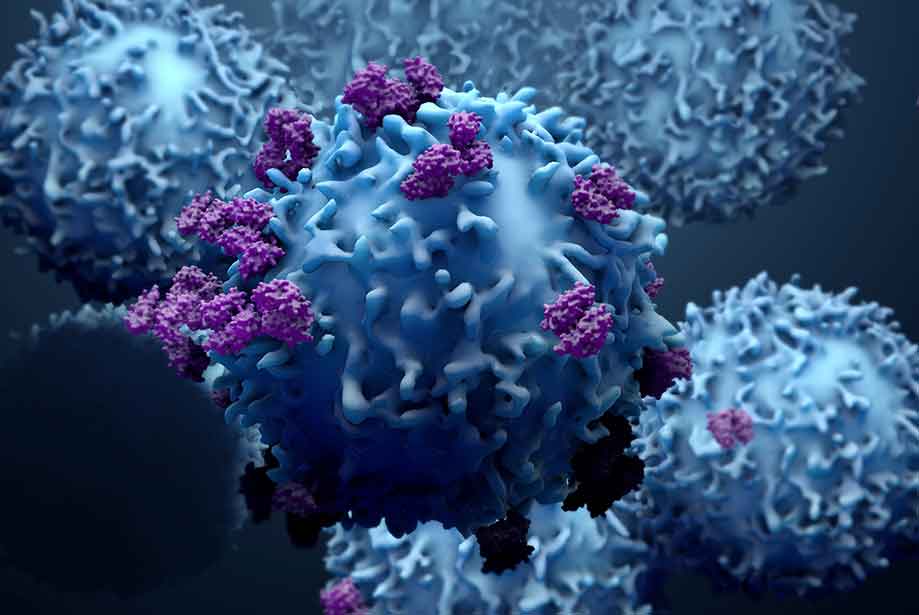
Immuno-oncology, also known as cancer immunotherapy, is an emerging sub-category of cancer research dedicated to examining ways in which the body’s immune system can be leveraged to fight cancer. Cancer cells that form solid tumors with microenvironments do so by initially evading destruction from host immunological defenses and surveillance long enough to colonize and accumulate in size and cell number before further expanding. Understanding how cancer cells interact with immune cells to survive offers great potential for more targeted treatments. The ultimate aim for researchers in this field is to develop immunotherapies that are more specific for cancers and less costly to endure by patients than traditional nonsurgical treatments. Traditional treatments such as chemotherapy, radiation or drugs that inhibit angiogenesis are not always specific enough to eliminate just the cancerous cells and may also target normal cells.
Therefore, a primary research focus has been to understand how immune checkpoint proteins operate to regulate the immune response. Most immune checkpoints exert their excitatory or inhibitory effects through membrane-bound receptor-ligand interactions. Cancer cells can manipulate these interactions to avoid detection and destruction by effectors of the innate and/or adaptive immune response. Advancements in this area of immuno-oncology research have facilitated the design of compounds or antibodies to target these receptors or ligands and block their functionality—these are known as immune checkpoint inhibitors (ICIs). At Enzo, we offer a complete set of solutions for
Immuno-oncology research focused on immune checkpoints including antibodies, ELISA kits, recombinant proteins and reagents for flow cytometry to help measure the magnitude of immune responses in tissues after screening ICIs. Our diverse scientific and technical expertise has helped us create innovative assays and reagents to help support cancer research. We provide flexible and validated tools to help you generate reliable and consistent results.
What are Immune Checkpoints?
Immune checkpoints manage the flow of co-stimulatory and inhibitory signals to regulate the magnitude and longevity of the immune response. Checkpoints exert control over the extent of immune-activation or -suppression based on how and when these “on” or “off” signals are passed along in cellular pathways after the immune response is initiated. These checkpoints are vital for maintaining immunological homeostasis by promoting self-tolerance and protecting hosts from inflammatory tissue damage when the immune system is activated. As previously mentioned, cancer cells manipulate interactions between immune checkpoints and their ligands to survive. One strategy has been by expressing the negative regulators of these checkpoints to evade immune effector cells (NK Cells and T-Cells) from carrying out full-fledged anti-tumor immune responses and promote immunosuppression instead to facilitate solid tumor development within a host. Below we review some key checkpoint regulators with immuno-suppressive functions that are attractive targets for immuno-oncology research.
CTLA-4, PD-1, LAG-3, TIM-3- Key Immunosuppressive Checkpoints
The most studied negative regulator of immune checkpoints is Cytotoxic T-Lymphocyte Antigen 4 (CTLA-4 or CD152). CTLA-4 is a transmembrane glyco-protein receptor, a part of the immunoglobulin superfamily that suppresses the immune response by inducing inhibitory effects on activated T-cells. This immune checkpoint protein exerts inhibition on activated T cells by out-competing its homologous “on” co-stimulatory immune signal, CD28, for binding spots to CD80/CD86 (B7 protein) presented as cognate antigen by antigen presenting cells (APCs) to turn off T-cell activation. To begin the T-cell immune response, T-cells are activated by APCs presenting Major Histocompatibility Complex I (MHC I) as cognate antigen as a primary signal to partially activate T-cells. Then, to fully activate T-cells and recruit additional effector cells, B7 protein is presented by APCs to T-cells expressing co-stimulatory CD28 and binding results in kinase activity that transmits T-cell signal downstream to amplify the initial signal. Both signals together drive T-cells to secrete Interleukin-2 (IL-2) which stimulates T-cell proliferation. CTLA-4 is able to outcompete CD28 because it has greater binding affinity to B7 making it effective in turning off T-cell activation. Upon CTLA-4 binding to B7 on APCs, phosphorylation of CTLA-4 by phosphoinositide 3-kinase (PI3K) results in the subsequent activation of phosphatases, SHP2 and PP2A, which counteract kinase signals looking to amplify T-signal produced by CD28 binding to B7 and reducing T-cell involvement and proliferative activities from lymphoid organs.
Another key negative regulator of immune checkpoints is a transmembrane protein from the CD28/CTLA-4 family known as Programmed Death-1 (PD-1). It enacts its immuno-suppressive function through binding to its associated Programmed Death Ligands PD-L1 (B7-H1 or CD274) and PD-L2 (B7-DC or CD273). PD-1 receptors and their ligands are expressed on the surfaces of mature T-cells, immunosuppressive regulatory T cells (Tregs) and other immune cells (macrophages, monocytes, B-cells, APCs, NK cells). Binding of PD-1 to either PD ligand results in inhibiting T-cell proliferation and effector functions (cytokine production and release). Initially, PD-1 activation causes the dephosphorylation of T-cell kinase ZAP70 and PI3K which prevents T-cell signal cascades from being passed downstream for AKT/mTOR pathway activation. The lack of AKT signaling leads to decreased Tc cell proliferation and production of inflammatory cytokines by Cytotoxic T Cells. Tumor cells express their own PD-L1 to take advantage of immune-subversion by this inhibitory immune checkpoint regulator to diminish T-cell activation and avoid destruction.

Figure 1: IHC using anti-PD-L1 (Prod. No. ENZ-ABS693) on lung adenocarcinoma tissue.
|
Lymphocyte-activation gene 3 (LAG-3 or CD233) is an immune checkpoint protein that belongs to the immunoglobulin superfamily. It is a structural homolog to CD4 that binds to major histocompatibility complex (MHC) class II molecules on APCs. LAG-3 is expressed on NK cells, B cells, T cells and dendritic cells. The signaling mechanism of LAG-3 is still largely unknown but it exerts its immuno-suppressive effect by enhancing Tregs activity and dampening T-cell signaling to prevent CD4+ and CD8+ T cell activation which reduces cytokine production and T-cell proliferation. LAG-3 is immuno-inhibitory much like CTLA-4 and PD-1 but promotes a less potent immunosuppressive effect on T-cells.
T-cell immunoglobulin mucin 3 (TIM-3) is an immune checkpoint protein expressed on cell surfaces of CD4+ and CD8+ T cells and other immune effector cells. It binds to its primary ligand galectin-9 to exert its immuno-suppressive function on T cells. Receptor-ligand binding causes cytoplasmic protein Bat3 which is already bound to TIM-3 to dissociate which disrupts interferon-y production and T-cell proliferation.
Enzo Life Sciences offers a comprehensive
Immuno-oncology research portfolio. At Enzo, we believe in providing scientists with novel tools that will help support cancer discovery. We have our
SCREEN-WELL® Cancer Library, a collection of 275 compounds that can be used for screening checkpoint inhibitors. Furthermore, our protein portfolio includes
PD-1,
PDL-1,
IL-2,
SHP-2 and
MEGACD40L® to aid your research. We offer
CTLA-4,
TIM-3,
PD-1,
PD-L1 antibodies and many more. Our growing list of over
1000 IHC validated antibodies includes those for the detection of key signaling proteins, cell surface markers, and mediators of cell death targeted in this research area. You can use our antibodies through our
Worry-Free Antibody Trial Program. Please check out our
Immuno-oncology platform for more information or contact our
Technical Support Team for further assistance.

 (ALX-804-806) on human tonsil tissue")


