Cytogenetics describes the study of chromosome structure. The earliest cytogenetic studies involved visualization and counting of whole chromosomes. Chromosome analysis is now used to detect genomic modifications and their role in disease. Additional or missing chromosomes can cause severe diseases. One of the most famous examples is an additional chromosome 21, which is associated with a condition called trisomy 21, better known as Down syndrome. Obvious translocations can also be detected by this method. The “Philadelphia chromosome”, which is linked to chronic myelogenous leukemia (CML), is the result of a translocation between chromosome 22 and 9. Specifically, a part of chromosome 22 translocates to chromosome 9, forming the BCR-ABL fusion gene. ABL1 has an increased kinase activity when it is fused to BCR, leading to CML.
Identification of Insertions, Deletions and Translocations
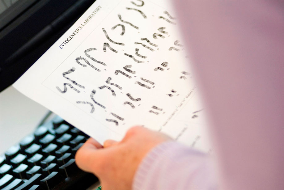
Chromosomes can be stained with the Giemsa stain to visualize G-bands. Each chromosome has a characteristic G-banding pattern and comparison of these patterns between two sample cells makes it possible to detect abnormalities, such as insertions, deletions and translocations. The disadvantage of this method is that only metaphase chromosomes can be stained with Giemsa. As metaphase chromosomes are highly condensed, high resolution results are not possible.
More Advanced Methods to Study Chromosome and Gene Modifications
FISH
One advanced way to detect insertions, deletions, duplications and translocations are probe-based methods. A widely used method is fluorescence in-situ hybridization (FISH). Probes that are complementary to gene sequences of interest in the genome are fluorescently labeled. These probes bind to the parts of the chromosome where this sequence is located. Duplication of a sequence would lead to an additional fluorescent spot, translocation would move the spot away from its usual site. FISH probes can hybridize to metaphase spreads and also to interphase chromosomes, allowing a much higher resolution than G-banding karyotyping.
FISH probes can be prepared by nick translation, which requires DNA Polymerase I and DNase I. DNase I introduces random nicks in the template DNA and DNA Polymerase I extends the 3’-OH end of the nick with nucleotide residues, thereby eliminating residues from the 5’-PO4 end of the nicks. In the presence of fluorescently labeled nucleotides, fluorescently labeled DNA that can then be used as a probe is generated. Advantages of preparing FISH probes rather than using commercially available probes are (1) the flexibility in preparing probes for any target sequence of interest and (2) the price. Enzo Life Sciences offers the Nick translation DNA labeling system 2.0, which enables the user to generate FISH probes from template DNA containing the sequence of interest. Instead of fluorophore-labeled probes, digoxigenin or biotin-labeled probes can be prepared, if desired. This allows chromogenic signal detection, which is widely used in immunohistochemistry (IHC). Chromogenic in situ hybridization (CISH) is preferred in diagnostic labs, as the signal can be easily detected with bright-field microscopes.
CGH
An even more advanced method to analyze chromosome and gene modifications is comparative genomic hybridization (CGH), which can be used for genome-wide analysis. In its original form, DNA is isolated and fragmented from an experimental sample and a control sample. Both samples are then labeled with two different fluorescent colors, usually green and red. The DNA samples are mixed and the generated probes compete for hybridization on a normal chromosome. If both samples are identical, a mixed yellow/orange color is seen everywhere. A red signal indicates an overrepresentation of the red-labeled experimental DNA, the result of gene duplication. A green signal, on the other hand, indicates a gene loss.
This form of CGH requires metaphase chromosomes, which reduces the resolution. More recently, array-based CGH (aCGH) evolved. In aCGH, thousands of DNA probes that consist of known sequences in the genome are attached to a microchip. As with standard CGH, two DNA samples are labeled in two different colors and then loaded onto the chip for competitive hybridization. aCGH allows the identification of gene or sequence deletions and amplifications in a very precise manner. In one single experiment, many thousand sequences are analyzed simultaneously.
Enzo Life Sciences offers labeling kits for aCGH. They are based on a proprietary labeling technology that leads to a high labeled DNA yield and a high specific activity, resulting in very low DLR scores and clear and reliable results. The
CYTAG® TotalCGH Labeling kit allows the realization of single nucleotide polymorphism (SNP) arrays in addition to standard CGH arrays. For a SNP array, the DNA has to be digested with restriction enzymes prior to labeling. The labeled DNA probes are then hybridized to special SNP microchips that contain probes that detect known SNPs in the genome. Our
CYTAG® SuperCGH Labeling kit is specifically designed for low input samples as low as 50ng.
FISH or aCGH?
In FISH, multiple colored probes can be hybridized to the experimental sample, but the number of different probes is limited to the number of different colors that are available. aCGH however allows for easy analysis of thousands of sequences with a very high resolution and moreover makes the detection of SNPs possible. Both methods can be used in a complimentary way to confirm a result with two different methods. In Summary, both FISH and aCGH are very powerful tools to study genomic alterations.
Check out our
aCGH platform and nick translation to prepare
FISH probes guide for further information or contact our
Technical Support Team for further assistance.