Replaces Prod. #: ENZ-51038
-
Provides a sensitive cell-based assay of drug responsiveness to identify inhibitors relevant to neurodegenerative disease in an authentic cellular context
-
Reliable and simple assay doesn’t require non-physiological protein mutations or genetically engineered cell lines
-
Validated under a wide range of conditions and with small molecule modulators demonstrating suitability for screening compounds of potential therapeutic value
-
Fixed-cell assay is optimized for antibody co-localization studies
-
Easily quantifies aggresome and related inclusion bodies by flow cytometry
-
Useful for the study of neurodegenerative diseases, liver disease, toxicology studies and much more
Aggresomes are inclusion bodies of aggregated, misfolded proteins that form when the ubiquitin-proteasome protein-degradation machinery is overwhelmed. Typically, aggresomes form in response to cellular stress and provide a cytoprotective mechanism by isolating misfolded proteins, leading ultimately to their clearance by autophagy. Protein aggregate formation is furthermore a hallmark of a variety of human diseases, such as Alzheimer's, Parkinson's, Amyotrophic lateral sclerosis or alcoholic liver disease.
The PROTEOSTAT® Aggresome Detection Reagent contained in this kit is a molecular rotor dye. While in solution, free intramolecular rotation along a single central bond prevents fluorescence. The PROTEOSTAT® dye specifically intercalates into the cross-beta spine of quaternary protein structures typically found in misfolded and aggregated proteins, which will inhibit the dye’s rotation and lead to a strong fluorescence.
PROTEOSTAT® Aggresome Detection kit provides a rapid, specific and quantitative approach to label aggresomes in cells, eliminating the need of artificial protein mutations. PROTEOSTAT® dye has been validated under a wide range of conditions and with small molecule modulators, demonstrating its suitability for compound screening. Furthermore, it is suitable for multiplex co-immunofluorescence to study your target of interest in the context of autophagy and aggresome formation.
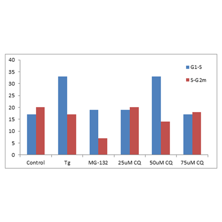
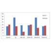
K562 cells were treated with reagents to induce ER stress and incomplete autophagy using Thapsigargin (Tg, 0.1 µM), 25, 50 and 75 µM Chloroquine (CQ) for 24 h respectively. A positive control was employed using the proteasome inhibitor, MG-132 (5 µM) for 24 h. Pelleted cells were fixed and permeabilized. Cells were then labelled for 30 min at RT with 300 µl PROTEOSTAT® Aggresome Detection reagent (Enzo Life Sciences) according to the manufacturer’s instructions and 1 µg/ml DAPI (for cell cycle determination). Relative increase in Aggresome Propensity Factor (APF) of S phase above that of G1 and that of G2m above S phase was determined for each treatment and compared to control values. ER stress inducer, Tg and 50 µM CQ was shown to both up-regulate Aggresomes more in S phase than G1 compared to controls. While proteasome inhibitor, MG-132 and 50 µM CQ was shown to down-regulate Aggresomes more in G2m than S phase than control levels.Courtesy of the Flow Cytometry Core Facility, Blizard Institute, Queen Mary University of London, London, UK.
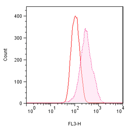
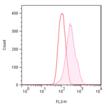
Flow cytometry-based analysis. Jurkat cells were mock-induced with 0.2% DMSO or induced with 5 µM MG-132 overnight at 37°C. After treatment, cells were fixed and incubated with PROTEOSTAT® dye, then analyzed by flow cytometry without washing using a 488 nm laser in the FL3 channel. In MG-132 treated cells, fluorescent red signal increases about 3-fold. The described assay allows assessment of the effects of protein aggregation.
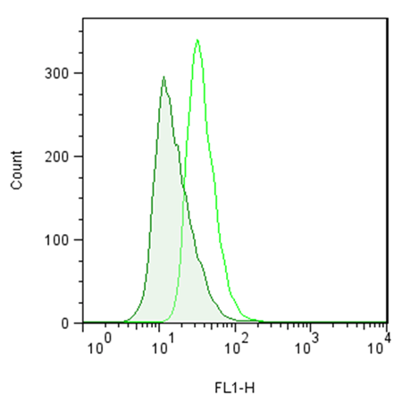
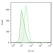
Flow cytometry-based analysis. Jurkat cells were mock-induced with 0.2% DMSO or induced with 5 µM MG-132 overnight at 37°C. After treatment, cells were fixed and incubated with Fluorescein-p62 Ab (1:500 dilution of stock), then after washing, analyzed by flow cytometry using a 488 nm laser in the FL1 channel. In MG-132 treated cells, Fluorescein-p62 Ab signal increases about 2.5-fold.
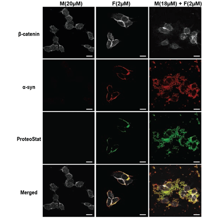
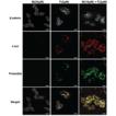
α-syn mixture forms aggregates at the cell plasma membrane that are internalized into the M17 neuroblastoma cell line. M17 cells were treated with α-syn monomers (M), sonicated α-syn PFFs (F) or α-syn mixture (M+F). After 4 days, M17 cells were washed 3 times prior to fixation and then stained with the PROTEOSTAT® aggregation dye (green) and immunostained against α-syn (red) and β-catenin (grey). Scale bars = 20 µm. Image used with permission from authors from “Fibril growth and seeding capacity play key roles in α-synuclein-mediated apoptotic cell death. A-L Mahul-Mellier, et al.; Cell Death & Differentiation (2015).” (doi:10.1038/cdd.2015.79).
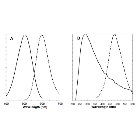
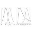
Emission/Excitation Spectra of PROTEOSTAT® Dye and Nuclear Stain
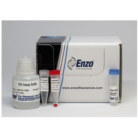
ProteoStat® Aggresome Detection Kit
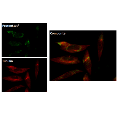
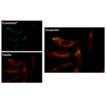
Aggresomes with HeLa cells, previously treated for 12 hours with 5µM MG-132, detected by ProteoStat® Aggresome dye (upper left, appearing here in green) showing co-localization with Alexa Fluor® 647-Tubulin antibody (lower left, appearing in red) and composite image (right), as observed by fluorescence microscopy.
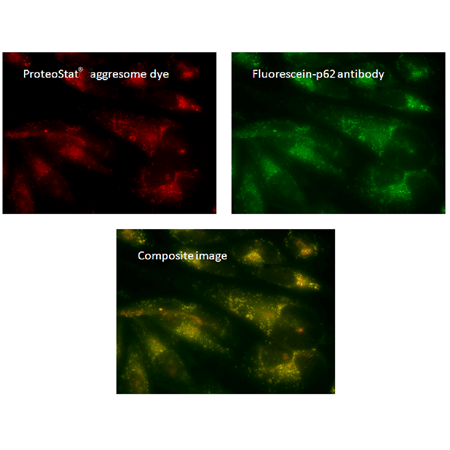
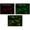
Aggresomes within HeLa cells, previously treated for 12 hours with 5μM MG-132, detected by ProteoStat® aggresome dye (upper left) showing co-localization with fluorescein-p62 antibody (upper right) and composite image (lower center), as observed by fluorescence microscopy.
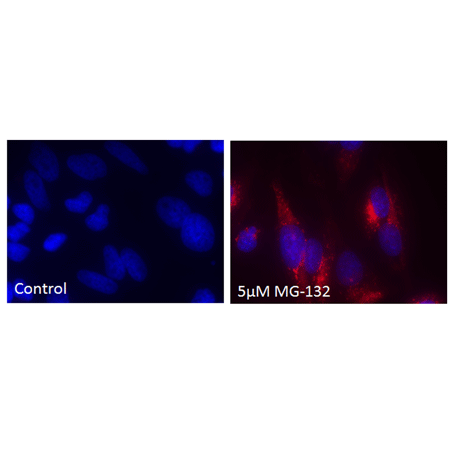
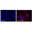
Aggresomes with HeLa cells, treated for 12 hours with 5µM MG-132 (right), detected by ProteoStat® Aggresome dye (red) and counterstained with Hoechst 33342. Control panel on left is untreated.
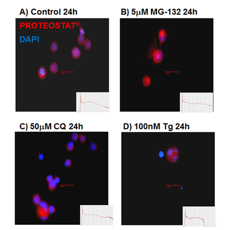
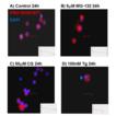
Aggresomes and aggresome-like inclusion bodies (ALIS) produced by ER stress related autophagy were imaged by epi-fluorescence microscopy. K562 cells were A) untreated or treated with B) 5 mM MG-132 C) 50 mM CQ or D) 100 nM thapsigargin (Tg) for 24 h. Cells were then fixed and permeabilised and incubated with PROTEOSTAT® dye (1:10,000 dilution) for 30 min at RT. PROTEOSTAT® staining indicating aggresomes & ALIS are shown in red with DAPI staining shown in blue. Courtesy of the Flow Cytometry Core Facility, Blizard Institute, Queen Mary University of London, London, UK.
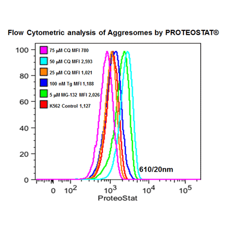
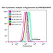
Flow cytometric analysis of protein aggresomes induced by ER stress related autophagy. K562 cells were untreated or treated with 25, 50, 75 mM CQ, 100 nM thapsigargin (Tg) or 5 mM MG-132 for 24 h. Cells were then fixed and permeabilised and incubated with PROTEOSTAT® dye (1:10,000 dilution) for 30 min at RT. Cells (30,000) were then analysed on the blue 610/20nm channel of a BD LSRII. Courtesy of the Flow Cytometry Core Facility, Blizard Institute, Queen Mary University of London, London, UK.
Please mouse over
Product Details
| Applications: | Flow Cytometry, Fluorescence microscopy, Fluorescent detection
|
| |
| Application Notes: | The PROTEOSTAT® Aggresome detection kit assay is potentially applicable to the identification of small molecules that inhibit aggresome formation as well as immuno-localization studies between aggregated protein cargo and the various proteins implicated in aggresome formation, such as histone deacetylase 6, parkin, ataxin-3, dynein motor complex and ubiquilin-1. |
| |
| Quality Control: | A sample from each lot of PROTEOSTAT® Aggresome detection kit is used to stain Jurkat cells and analyzed by flow cytometry, using the procedures described in the user manual. The aggresome activity factor (AAF) values for the samples are greater than 25. |
| |
| Quantity: | For ENZ-51035-0025:
25 flow cytometry assays or 50 microscopy assays
For ENZ-51035-K100:
100 flow cytometry assays or 200 microscopy assays |
| |
| Use/Stability: | With proper storage, the kit components are stable for one year from date of receipt. |
| |
| Handling: | Protect from light. Avoid freeze/thaw cycles. |
| |
| Shipping: | Blue Ice |
| |
| Short Term Storage: | -20°C |
| |
| Long Term Storage: | -80°C |
| |
| Contents: | PROTEOSTAT®Aggresome detection reagent
Hoechst 33342 Nuclear stain
Proteasome Inhibitor (MG-132)
10X Assay Buffer |
| |
| Technical Info/Product Notes: | The PROTEOSTAT® Aggresome Detection Kit is a member of the CELLESTIAL® product line, reagents and assay kits comprising fluorescent molecular probes that have been extensively benchmarked for live cell analysis applications. CELLESTIAL® reagents and kits are optimal for use in demanding imaging applications, such as confocal microscopy, flow cytometry and HCS, where consistency and reproducibility are required.
Featured In:
High-throughput image-based aggresome quantification (SLAS Discov 2020; 25:783-791).
Application: Optimized to monitor aggresome formation in a miniaturized, automated, and quantitative phenotypic assay. The approach is validated by screening a chemical library of 1280 compounds.
Application Note:
A Red-emitting Fluorescent Probe for Rapid Detection of Protein and Peptide Aggregates in Post-mortem Human Brain Tissue Sections from Patients Diagnosed with Alzheimer’s and Parkinson’s Disease
Monitoring the Accumulation and Clearance of Exogenously Introduced Beta-amyloid in a Cell-Based Model of Alzheimer’s Disease by Fluorescence Microscopy and Fluorescence Microplate Assay
Towards Understanding the Molecular Basis of Parkinson’s Disease: Cell-based Model of Mitophagy and Aggresome Accumulation
Detection of bacterial aggregation by flow cytometry
Cell-Based Screening of Focused Bioactive Compound Libraries: Assessing Small Molecule Modulators of the Canonical Wnt Signaling and Autophagy-Lysosome Pathways
Cited samples:
For an overview on cited samples please click here.
Enzo and PROTEOSTAT® are trademarks of Enzo Life Sciences, Inc. Several of Enzo’s products and product applications are covered by US and foreign patents and patents pending. |
| |
| Regulatory Status: | RUO - Research Use Only |
| |
Product Literature References
Immunofluorescence and Aggresome Staining of Nothobranchius furzeri Cryosections: S. Bagnoli, et al.; Cold Spring Harb. Protoc. (2023),
Abstract;
Immunofluorescence and Aggresome Staining of Nothobranchius furzeri Cryosections: S. Bagnoli, et al.; Cold Spring Harb. Protoc.
2023, 693 (2023),
Abstract;
Insulin regulates human pancreatic endocrine cell differentiation in vitro: P. Cota, et al.; Mol. Metab.
79, 101853 (2023),
Abstract;
Oxidative-Stress-Mediated ER Stress Is Involved in Regulating Manoalide-Induced Antiproliferation in Oral Cancer Cells: S.Y. Peng, et al.; Int. J. Mol. Sci.
24, 3987 (2023),
Abstract;
Transcriptomic characterization of Lonrf1 at the single-cell level under pathophysiological conditions: D. Li, et al.; J. Biochem.
173, 459 (2023),
Abstract;
A meiotic switch in lysosome activity supports spermatocyte development in young flies but collapses with age: T.J. Butsch, et. al.; iScience
25, 104382 (2022),
Abstract;
A novel HIF-2α targeted inhibitor suppresses hypoxia-induced breast cancer stemness via SOD2-mtROS-PDI/GPR78-UPRER axis: Y. Yan, et al.; Cell Death Differ.
1038, s41418 (2022),
Abstract;
Apoptotic mechanism in human brain microvascular endothelial cells triggered by 4′-iodo-α-pyrrolidinononanophenone: Contribution of decrease in antioxidant properties: Y. Sakai, et al.; Toxicol. Lett.
351, 127 (2022),
Abstract;
Calcium dysregulation potentiates wild-type myocilin misfolding: implications for glaucoma pathogenesis: E.G. Saccuzzo, et al.; J. Biol. Inorg. Chem.
27, 553 (2022),
Abstract;
Copper oxide nanoparticles trigger macrophage cell death with misfolding of Cu/Zn superoxide dismutase 1 (SOD1): G. Gupta, et al.; Part. Fibre Toxicol.
19, 33 (2022),
Abstract;
De novo sphingolipid biosynthesis necessitates detoxification in cancer cells: M.E. Spears, et al.; Cell Rep.
40, 111415 (2022),
Abstract;
Engineered Riboswitch Nanocarriers as a Possible Disease-Modifying Treatment for Metabolic Disorders: S. Zilberzwige-Tal , et al.; ACS Nano
16, 11733 (2022),
Application(s): Quantification of amyloid protein in yeast after treatment with riboswitch nanocarriers,
Abstract;
Error-prone protein synthesis recapitulates early symptoms of Alzheimer disease in aging mice: M. Brilkova, et al.; Cell Rep.
40, 111433 (2022),
Abstract;
Human Cathelicidin Peptide LL-37 Induces Cell Death in Autophagy Dysfunctional Endothelial Cells: K. Suzuki, et al.; J. Immunol.
208, 2163 (2022),
Abstract;
Identification of Novel Oxindole Compounds That Suppress ER Stress-Induced Cell Death as Chemical Chaperones: Y. Hasegawa, et al.; ACS Chem. Neurosci.
13, 1055 (2022),
Abstract;
IGF-1 as a Potential Therapy for Spinocerebellar Ataxia Type 3: Y.-S. Lin, et al.; Biomedicines
10, 505 (2022),
Abstract;
Induction of Oxidative Stress in SH-SY5Y Cells by Overexpression of hTau40 and Its Mitigation by Redox-Active Nanoparticles: N. Pieńkowska, et al.; Int. J. Mol. Sci.
24, 359 (2022),
Abstract;
New lessons on TDP-43 from old N. furzeri killifish: A. Louka, et al.; Aging Cell
21, e13517 (2022),
Abstract;
Premature aging in mice with error-prone protein synthesis: D. Shcherbakov, et al.; Sci. Adv.
8, eabl9051 (2022),
Abstract;
Proteasome inhibition triggers the formation of TRAIL receptor 2 platforms for caspase-8 activation that accumulate in the cytosol: C.T. Hellwig, et al.; Cell Death Differ.
29, 147 (2022),
Abstract;
Quantification of noradrenergic-, dopaminergic-, and tectal neurons during aging in the short-lived killifish Nothobranchius furzeri: S. Bagnoli, et al.; Aging Cell
21, e13689 (2022),
Abstract;
The Nrf1 transcription factor is induced by patulin and protects against patulin cytotoxicity: J.J.W. Han, et al.; Toxicology
471, 153173 (2022),
Abstract;
αA and αB peptides from human cataractous lenses show antichaperone activity and enhance aggregation of lens proteins: O. Srivastava, et al.; Mol. Vis.
28, 147 (2022),
Abstract;
Artepillin C, a major component of Brazilian green propolis, inhibits endoplasmic reticulum stress and protein aggregation: Y. Hirata, et al.; Eur. J. Pharmacol.
912, 174572 (2021),
Abstract;
ATP-citrate lyase promotes axonal transport across species: A. Even, et al.; Nat. Commun.
12, 5878 (2021),
Abstract;
Cell-Instructive Surface Gradients of Photoresponsive Amyloid-like Fibrils: A.M. Bender, et al.; ACS Biomater. Sci. Eng.
7, 4798 (2021),
Abstract;
Dual Targeting of Endoplasmic Reticulum by Redox-Deubiquitination Regulation for Cancer Therapy: B. Cai, etr al.; Int. J. Nanomedicine
16, 5193 (2021),
Abstract;
Inhibition of cardiac PERK signaling promotes peripartum cardiac dysfunction: T. Shimizu, et al.; Sci. Rep.
11, 18687 (2021),
Abstract;
New evidences of ubiquitin-proteasome system activity in human sperm: J.V. Silva, et al.; Biochim. Biophys. Acta Mol. Cell. Res.
1868, 118932 (2021),
Application(s): Microscopy; detection of aggresome-like inclusion bodies in human sperm,
Abstract;
Novel blood test for early biomarkers of preeclampsia and Alzheimer’s disease: S. Cheng, et al.; Sci. Rep.
11, 15934 (2021),
Abstract;
Proteotoxic stress is a driver of the loser status and of cell competition: M. E. Baumgartner, et al.; Nat. Cell Biol.
23, 136 (2021),
Application(s): Fluorescence imaging of fly larvae,
Abstract;
Full Text
Sodium sulfite causes gastric mucosal cell death by inducing oxidative stress: O. Moeri, et al.; Free Radic. Res.
55, 731 (2021),
Abstract;
The intrinsic chaperone network of Arabidopsis stem cells confers protection against proteotoxic stress: E. Llamas, et al.; Aging Cell
20, e13446 (2021),
Application(s): Quantitative fluorescence imaging of Arabidopsis roots,
Abstract;
Full Text
The mechanism of cell death induced by silver nanoparticles is distinct from silver cations: M.M. Rhode, et al.; Part. Fibre Toxicol.
18, 37 (2021),
Abstract;
4-Phenylbutyrate ameliorates apoptotic neural cell death in Down syndrome by reducing protein aggregates: K. Hirata, et al.; Sci. Rep.
10, 14047 (2020),
Abstract;
Full Text
Cytokinin-induced protein synthesis suppresses growth and osmotic stress tolerance: S.S. Karunadasa, et al.; New Phytol.
227, 50 (2020),
Application(s): Confocal microscopy; A. thaliana seedlings,
Abstract;
Differentiation Drives Widespread Rewiring of the Neural Stem Cell Chaperone Network: W.I.M. Vonk, et al.; Mol. Cell
78, 328 (2020),
Abstract;
DMSO impairs the transcriptional program for maternal-to-embryonic transition by altering histone acetylation: M.H. Kang, et al.; Biomaterials
230, 119604 (2020),
Application(s): Confocal microscopy using mouse embryos,
Abstract;
Downregulated miR-18b-5p triggers apoptosis by inhibition of calcium signaling and neuronal cell differentiation in transgenic SOD1 (G93A) mice and SOD1 (G17S and G86S) ALS patients: K.Y. Kim, et al.; Transl. Neurodegener.
9, 23 (2020),
Abstract;
Full Text
Galectin-3 promotes Aβ oligomerization and Aβ toxicity in a mouse model of Alzheimer’s disease: C.C. Tao, et al.; Cell Death Differ.
27, 192 (2020),
Application(s): Visualisation of amyloid plaques on mouse brain sections,
Abstract;
Full Text
hnRNPDL phase separation is regulated by alternative splicing and disease-causing mutations accelerate its aggregation: C. Batlle, et al.; Cell Rep.
30, 1117 (2020),
Application(s): Fluorescence microscopy using aggregated proteins in solution,
Abstract;
Intravaginal poly-(D, L-lactic-co-glycolic acid)-(polyethylene glycol) drug-delivery nanoparticles induce pro-inflammatory responses with Candida albicans infection in a mouse model: T.T. Lina, et al.; PLoS One
15, e0240789 (2020),
Abstract;
Full Text
Keap1 governs ageing-induced protein aggregation in endothelial cells: A. Kopacz,et al.; Redox Biol.
34, 101572 (2020),
Abstract;
Mitochondrial dysfunction generates aggregates that resist lysosomal degradation in human breast cancer cells: T. G. Biel, et al.; Cell Death Dis.
11, 460 (2020),
Application(s): Fluorescence Imaging and Flow Cytometry of human breast cancer cells MDA-MB-231,
Abstract;
Full Text
Relationship between the Length of Sperm Tail Mitochondrial Sheath and Fertility Traits in Boars Used for Artificial Insemination: K. Kerns, et al. ; Antioxidants (Basel)
9, E1033 (2020),
Application(s): Microscope imaging-morphometry and image-based flow cytometry on sperm samples,
Abstract;
Targeting p53 and histone methyltransferases restores exhausted CD8+ T cells in HCV infection: V. Barili, et al.; Nat. Commun.
11, 604 (2020),
Application(s): Flow cytometry using human PBMCs,
Abstract;
Full Text
West Nile virus capsid protein inhibits autophagy by AMP-activated protein kinase degradation in neurological disease development: S. Kobayashi, et al.; PLoS Pathog.
16, e1008238 (2020),
Application(s): Confocal and fluorescence microscopy using SK-N-SH cells,
Abstract;
A Flow Cytometric Study of ER Stress and Autophagy: A. Popat, et al.; Cytometry A
95, 672 (2019),
Abstract;
Full Text
A new histone deacetylase inhibitor enhances radiation sensitivity through the induction of misfolded protein aggregation and autophagy in triple-negative breast: H.W. Chiu, et al.; Cancers (Basel)
11, 1703 (2019),
Application(s): Fluorescence microscopy using 4T1, MDA-MB-231, and MCF-10A cells,
Abstract;
Full Text
An SOD1 deficiency aggravates proteasome inhibitor bortezomib-induced testicular damage in mice: T. Homma & J. Fujii; Biochim. Biophys. Acta Gen. Subj.
1863, 1108 (2019),
Application(s): Mouse cryosectioned tissue,
Abstract;
Autophagy augmentation alleviates cigarette smoke-induced CFTR-dysfunction, ceramide-accumulation and COPD-emphysema pathogenesis: M. Bodas, et al.; Free Radic. Biol. Med.
131, 81 (2019),
Application(s): Co-staining and fluorescence microscopy using human and murine lung tissue sections,
Abstract;
Bassoon proteinopathy drives neurodegeneration in multiple sclerosis: B. Schattling, et al.; Nat. Neurosci.
22, 887 (2019),
Application(s): Confocal microscopy using mouse neurons,
Abstract;
Cardiac-specific Mst1 deficiency inhibits ROS-mediated JNK signalling to alleviate Ang II-induced cardiomyocyte apoptosis: Z. Cheng, et al.; J. Cell. Mol. Med.
23, 543 (2019),
Abstract;
Full Text
CBP/p300 Bromodomains Regulate Amyloid-like Protein Aggregation upon Aberrant LysineAcetylation: H. Olzscha, et al.; Cell Chem. Biol.
24, 9 (2019),
Abstract;
Cell-based HTS identifies a chemical chaperone for preventing ER protein aggregation and proteotoxicity: K. Kitakaze, et al.; eLife
8, e43302 (2019),
Application(s): Fluorescence microscopy and high-content analysis system using HEK293 cells,
Abstract;
Full Text
Designer amyloid cell-penetrating peptides for potential use as gene transfer vehicles: C. Kokotidou, et al.; Biomolecules
10, 7 (2019),
Application(s): Confocal microscopy using HEK293 cells,
Abstract;
Disturbances in H+ dynamics during environmental carcinogenesis: D. Lagadic-Gossmann, et al.; Biochimie
163, 171 (2019),
Application(s): Fluorescence microscopy using F258 cells,
Abstract;
Dual role of ribosome-binding domain of NAC as a potent suppressor of protein aggregation and aging-related proteinopathies: K. Shen, et al.; Mol. Cell
74, 729 (2019),
Application(s): Fluorescence microscopy using ST HDH Q7/7 and ST HDH Q7/111 cells,
Abstract;
Full Text
Elimination of protein aggregates prevents premature senescence in human trisomy 21 fibroblasts: N. Nawa, et al.; PLoS One
14, e0219592 (2019),
Abstract;
Full Text
Fibril formation and therapeutic targeting of amyloid-like structures in a yeast model of adenine accumulation: D. Laor, et al.; Neurochem. Res.
10, 62 (2019),
Application(s): Flow cytometry and confocal imaging of yeast cells,
Abstract;
Full Text
Functional transcriptome analysis in ARSACS KO cell model reveals a role of sacsin in autophagy: F. Morani, et al.; Sci. Rep.
9, 11878 (2019),
Application(s): Co-staining and fluorescence microscopy using SH-SY5Y cells,
Abstract;
Full Text
GRASP55 and UPR control interleukin-1β aggregation and secretion: M. Chiritoiu, et al.; Dev. Cell
49, 145 (2019),
Abstract;
Impaired mitochondrial calcium efflux contributes to disease progression in models of Alzheimer's disease: P. Jadiya, et al.; Nat. Commun.
10, 3885 (2019),
Application(s): Confocal microscopy using mouse neuronal cells,
Abstract;
Full Text
In vivo phenotyping of familial Parkinson's Disease with human induced pluripotent stem cells: a proof-of-concept study: O. Zygogianni, et al.; Neurochem. Res.
44, 1475 (2019),
Application(s): Co-staining and fluorescence microscopy,
Abstract;
Knockdown of TM9SF4 boosts ER stress to trigger cell death of chemoresistant breast cancer cells: Y. Zhu, et al.; Oncogene
38, 5778 (2019),
Application(s): Flow cytometry using MCF-7 cells,
Abstract;
Metformin augments panobinostat's anti-bladder cancer activity by activating AMP-activated protein kinase: K. Okubo, et al.; Transl. Oncol.
12, 669 (2019),
Application(s): Fluorescence microscopy using UMUC3 and J82 human bladder cancer cells,
Abstract;
Full Text
Monitoring the aggregation of GPCRs by fluorescence microscopy: S. Génier, et al.; Methods Mol. Biol.
1947, 289 (2019),
Application(s): Confocal microscopy,
Abstract;
mTORC1 signaling is palmitoylation-dependent in hippocampal neurons and non-neuronal cells and involves dynamic palmitoylation of LAMTOR1 and mTOR: S.S. Sanders, et al.; Front. Cell. Neurosci.
13, 115 (2019),
Application(s): Fluorescence microscopy using rat neurons,
Abstract;
Full Text
Rescue of tight junctional localization of a claudin-16 mutant D97S by antimalarial medicine primaquine in Madin-Darby canine kidney cells: K. Marukana, et al.; Sci. Rep.
9, 9647 (2019),
Application(s): Fluorescence microscopy using MDCK cells,
Abstract;
Full Text
Retinoic acid worsens ATG10-dependent autophagy impairment in TBK1-mutant hiPSC-derived motoneurons through SQSTM1/p62 accumulation: A. Catanese, et al.; Autophagy.
15, 1719 (2019),
Application(s): Fluorescence microscopy using tissue sections,
Abstract;
Screening Protein Aggregation in Cells Using Fluorescent Labels Coupled to Flow Cytometry: S. Ventura, et al.; Methods Mol. Biol.
1873, 195 (2019),
Application(s): Detailed Protocol for PROTEOSTAT analysis of protein aggregation in yeast and bacteria,
Abstract;
The center of olfactory bulb‐seeded α‐synucleinopathy is the limbic system and the ensuing pathology is higher in male than in female mice: D.M. Mason, et al.; Brain Pathol.
29, 741 (2019),
Application(s): Co-staining and fluorescence microscopy,
Abstract;
The chaperonin TRiC/CCT associates with Prefoldin through a conserved electrostatic interface essential for cellular proteostasis: D.Gestaut, et al.; Cell
177, 751 (2019),
Application(s): Fluorescence microscopy of yeast cells,
Abstract;
Full Text
TRPML1 promotes protein homeostasis in melanoma cells by negatively regulating MAPK and mTORC1 signaling: S.Y. Kasitinon, et al.; Cell Rep.
28, 2293 (2019),
Application(s): Fluorescence microscopy using melanoma cells,
Abstract;
Full Text
Acute exposure to organic and inorganic sources of copper: Differential response in intestinal cell lines: J. Keenan, et al.; Food Sci. Nutr.
6, 2499 (2018),
Application(s): Microplate assay using Caco-2 and HT-29 cells,
Abstract;
Full Text
ATF6 safeguards organelle homeostasis and cellular aging in human mesenchymal stem cells: S. Wang, et al.; Cell Discov.
4, 2 (2018),
Application(s): Flow cytometry using human mesenchymal stem cells,
Abstract;
Full Text
Cigarette smoke induced autophagy-impairment accelerates lung aging, COPD-emphysema exacerbations and pathogenesis: N. Vij, et al.; Am. J. Physiol. Cell. Physiol.
314, C73 (2018),
Abstract;
Culling less fit neurons protects against amyloid-β-induced brain damage and cognitive and motor decline: D.S. Coelho, et al.; Cell Rep.
25, 3661 (2018),
Application(s): Confocal microscopy using fly brain tissue section,
Abstract;
Full Text
Ethanol induced disordering of pancreatic acinar cell endoplasmic reticulum: an ER stress/defective unfolded protein response model: R.T. Waldron, et al.; Cell. Mol. Gastroenterol. Hepatol.
5, 479 (2018),
Application(s): Confocal microscopy using AR42J cells,
Abstract;
Full Text
iPSCs from a hibernator provide a platform for studying cold adaptation and its potential medical applications: J. Ou, et al.; Cell
173, 851 (2018),
Application(s): Fluorescence microscopy using squirrel neurons,
Abstract;
Full Text
Novel polyubiquitin imaging system, PolyUb-FC, reveals that K33-linked polyubiquitin is recruited by SQSTM1/p62: Y. Nibe, et al.; Autophagy
14, 347 (2018),
Application(s): Fluorescence microscopy using HEK293T cells,
Abstract;
Full Text
Spermine increases acetylation of tubulins and facilitates autophagic degradation of prion aggregates: K. Phadwal, et al.; Sci. Rep.
8, 10004 (2018),
Application(s): Confocal microscopy with CAD and SMB cells,
Abstract;
Full Text
The ubiquitin ligase UBR5 suppresses proteostasis collapse in pluripotent stem cells from Huntington's disease patients: S. Koyuncu, et al.; Nat. Commun.
9, 2886 (2018),
Application(s): Fluorescence microscopy using pluripotent stem cells,
Abstract;
Full Text
A leptospiral AAA+ chaperone-Ntn peptidase complex, HslUV, contributes to the intracellular survival of Leptospira interrogans in hosts and the transmission of leptospirosis: S.L. Dong, et al.; Emerg. Microbes Infect.
6, e105 (2017),
Application(s): Protein aggresome detection in intracellular leptospires,
Abstract;
Full Text
Augmentation of S-Nitrosoglutathione Controls Cigarette Smoke-Induced Inflammatory-Oxidative Stress and Chronic Obstructive Pulmonary Disease-Emphysema Pathogenesis by Restoring Cystic Fibrosis Transmembrane Conductance Regulator Function: M. Bodas, et al.; Antioxid. Redox Signal.
27, 433 (2017),
Application(s): Human and murine lung tissue,
Abstract;
Full Text
Characterization of cadmium chloride-induced BiP accumulation in Xenopus laevis A6 kidney epithelial cells: C.S. Shirriff, et al.; Comp. Biochem. Physiol. C. Toxicol. Pharmacol.
191, 117 (2017),
Application(s): Immunocytochemical analysis and laser scanning confocal microscopy,
Abstract;
Endoplasmic Reticulum Stress Response in Arabidopsis Roots: Y. Cho, et al.; Front. Plant Sci.
8, 144 (2017),
Application(s): Fluorescence Imaging of Arabidopsis roots,
Abstract;
Full Text
FKBP8 protects the heart from hemodynamic stress by preventing the accumulation of misfolded proteins and endoplasmic reticulum-associated apoptosis in mice: T. Misaka, et al.; J. Mol. Cell. Cardiol.
114, 93 (2017),
Abstract;
Histone deacetylase 6 (HDAC6) is an essential factor for oocyte maturation and asymmetric division in mice: D. Zhou, et al.; Sci. Rep.
7, 8131 (2017),
Application(s): Mouse Oocyte Samples,
Abstract;
Full Text
OSM mitigates post-infarction cardiac remodeling and dysfunction by up-regulating autophagy through Mst1 suppression: J. Hu, et al.; Biochim. Biophys. Acta
1863, 1951 (2017),
Abstract;
p53 amyloid formation leading to its loss of function: implications in cancer pathogenesis: S. Ghosh, et al.; Cell Death Differ.
24, 1784 (2017),
Application(s): p53 aggregate formation in SY5Y cells,
Abstract;
Full Text
Polyethylene glycol-functionalized poly (Lactic Acid-co-Glycolic Acid) and graphene oxide nanoparticles induce pro-inflammatory and apoptotic responses in Candida albicans-infected vaginal epithelial cells: R.D. Wagner, et al.; PLoS One
12, e0175250 (2017),
Abstract;
Full Text
Subcellular localization of the five members of the human steroid 5α-reductase family: A. Scaglione, et al.; Biochim. Open
4, 99 (2017),
Application(s): Fluorescence Microscopy of HeLa cells,
Abstract;
Full Text
Targeting mitochondrial dysfunction can restore antiviral activity of exhausted HBV-specific CD8 T cells in chronic hepatitis B: P. Fisicaro, et al.; Nat. Med.
23, 327 (2017),
Abstract;
A fast and specific method to screen for intracellular amyloid inhibitors using bacterial model systems: S. Navarro, et al.; Eur. J. Med. Chem.
121, 785 (2016),
Application(s): Confocal microscopy,
Abstract;
Ad-HGF improves the cardiac remodeling of rat following myocardial infarction by upregulating autophagy and necroptosis and inhibiting apoptosis: J. Liu, et al.; Am. J. Transl. Res.
8, 4605 (2016),
Abstract;
Full Text
Airway exposure to e-cigarette vapors impairs autophagy and induces aggresome formation: P.C. Shivalingappa, et al.; Antioxid. Redox Signal.
24, 186 (2016),
Abstract;
Bile acids protect expanding hematopoietic stem cells from unfolded protein stress in fetal liver: Y. Sigurdsson, et al.; Cell Stem Cell
18, 522 (2016),
Abstract;
Casitas B-cell lymphoma (Cbl) proteins protect mammary epithelial cells from proteotoxicity of active c-Src accumulation: C. Mukhopadhyay, et al.; PNAS
113, E8228 (2016),
Abstract;
Full Text
Comprehensive proteomic study of the antiproliferative activity of a polyphenol-enriched rosemary extract on colon cancer cells using nanoliquid chromatography-orbitrap MS/MS: A. Valdés, et al.; J. Proteome Res.
15, 1971 (2016),
Abstract;
Distinct roles for intracellular and extracellular lipids in hepatitis C virus infection: S. Narayanan, et al.; PLoS One
11, e0156996 (2016),
Application(s): Multiphoton microscopy using human hepatocellular carcinoma HuH7.5.1 cells,
Abstract;
Full Text
Lin28a protects against postinfarction myocardial remodeling and dysfunction through Sirt1 activation and autophagy enhancement: Y. Hao, et al.; Biochem. Biophys. Res. Commun.
479, 833 (2016),
Application(s): Aggresome and p62 detection,
Abstract;
LRRK2 interferes with aggresome formation for autophagic clearance: Y. Bang, et al.; Mol. Cell. Neurosci.
75, 71 (2016),
Application(s): Protein aggregate analysis,
Abstract;
Luteolin alleviates post-infarction cardiac dysfunction by up-regulating autophagy through Mst1 inhibition: J. Hu, et al.; J. Cell. Mol. Med.
20, 147 (2016),
Application(s): Confocal microscopy using mouse cardiomyocytes,
Abstract;
Full Text
Monitoring of dipeptidyl peptidase-IV (DPP-IV) activity in patients with mucopolysaccharidoses types I and II on enzyme replacement therapy - Results of a pilot study: K. Hetmanczyk, et al.; Clin. Biochem.
49, 458 (2016),
Application(s): Plasma DPP-IV enzyme assay,
Abstract;
Prophylactic neuroprotective efficiency of co-administration of Ginkgo biloba and Trifolium pretense against sodium arsenite-induced neurotoxicity and dementia in different regions of brain and spinal cord of rats: H.M. Abdou, et al.; Food Chem. Toxicol.
94, 112 (2016),
Application(s): Fluorescence microscopy on tissue sections,
Abstract;
Regulation of GPCR Expression through an interaction with CCT7, a subunit of the CCT/TRiC Complex: S. Genier, et al.; Mol. Biol. Cell
27, 3800 (2016),
Application(s): Immunofluorescence Staining and Confocal Microscopy, HEK 293 cells,
Abstract;
Full Text
Repression of the autophagic response sensitises lung cancer cells to radiation and chemotherapy: I.V. Karagounis, et al.; Br. J. Cancer
115, 312 (2016),
Application(s): Confocal microscopy using lung cancer cell lines,
Abstract;
Resveratrol reduces amyloid-beta (Aβ1-42)-induced paralysis through targeting proteostasis in an Alzheimer model of Caenorhabditis elegans: C. Regitz, et al.; Eur. J. Nutr.
55, 741 (2016),
Abstract;
Structure-based drug discovery for prion disease using a novel binding simulation: D. Ishibashi, et al.; EBioMedicine
9, 238 (2016),
Application(s): Confocal microscopy using mouse neuroblastoma Neuro 2a cells,
Abstract;
Synergistic myeloma cell death via novel intracellular activation of caspase-10-dependent apoptosis by carfilzomib and selinexor: S. Rosebeck, et al.; Mol. Cancer Ther.
15, 60 (2016),
Abstract;
Amyloidogenic lysozymes accumulate in the endoplasmic reticulum accompanied by the augmentation of ER stress signals: Y. Kamada, et al.; Biochim. Biophys. Acta
1850, 1107 (2015),
Application(s): Microscopy,
Abstract;
Characterization of aggregate/aggresome structures formed by polyhedrin of Bombyx mori nucleopolyhedrovirus: Z.J. Guo, et al.; Sci. Rep.
5, 14601 (2015),
Application(s): Fluorescence microscopy using Bombyx mori cell line BmN,
Abstract;
Full Text
Conophylline protects cells in cellular models of neurodegenerative diseases by inducing mammalian target of rapamycin (mTOR)-independent autophagy: Y. Sasazawa, et al.; J. Biol. Chem.
290, 6168 (2015),
Abstract;
Decreased proteasomal function accelerates cigarette smoke-induced pulmonary emphysema in mice: Y. Yamada, et al.; Lab. Invest.
95, 625 (2015),
Application(s): Aggresome detection by fluorescence microscopy in fibroblasts,
Abstract;
Defective autophagy is a key feature of cerebral cavernous malformations: S. Marchi, et al.; EMBO Mol. Med.
7, 1403 (2015),
Application(s): Aggresome detection in aggregated proteins and aggresome‐like inclusion bodies in fixed and permeabilized samples,
Abstract;
Full Text
Enhanced proteotoxic stress: one of the contributors for hyperthermic potentiation of the proteasome inhibitor bortezomib using magnetic nanoparticles: M.P. Alvarez-Berrios, et al.; Biomater. Sci.
3, 391 (2015),
Abstract;
Fibril growth and seeding capacity play key roles in α-synuclein-mediated apoptotic cell death: A.L. Mahul-Mellier, et al.; Cell Death Differ.
22, 2107 (2015),
Abstract;
In vitro administration of gold nanoparticles functionalized with MUC-1 protein fragment generates anticancer vaccine response via macrophage activation and polarization mechanism: T. Mocan, et al.; J. Cancer
6, 583 (2015),
Application(s): Aggresome detection by fluorescence microscopy in peritoneal macrophages,
Abstract;
Full Text
Intensified autophagy compromises the efficacy of radiotherapy against prostate cancer: M.I. Koukourakis, et al.; Biochem. Biophys. Res. Commun.
461, 268 (2015),
Application(s): Fluorescence microscopy ,
Abstract;
Mechanisms for autophagy modulation by isoprenoid biosynthetic pathway inhibitors in multiple myeloma cells: K.M. Dykstra, et al.; Oncotarget
6, 41535 (2015),
Application(s): Confocal microscopy using human myeloma cell lines,
Abstract;
Full Text
Mevalonate pathway regulates cell size homeostasis and proteostasis through autophagy: T.P. Miettinen, et al.; Cell Rep.
13, 2610 (2015),
Application(s): Flow cytometry analysis of protein aggregation using Jurkat, U2OS, Kc167 and HUVEC cells,
Abstract;
MiR-29b replacement inhibits proteasomes and disrupts aggresome+autophagosome formation to enhance the antimyeloma benefit of bortezomib: S. Jagannathan, et al.; Leukemia
29, 727 (2015),
Application(s): Detection of protein aggregates by fluorescence microscopy in multiple myeloma cell lines,
Abstract;
Full Text
Molecular chaperone GRP78 enhances aggresome delivery to autophagosomes to promote drug resistance in multiple myeloma: M.A. Abdel Malek, et al.; Oncotarget
6, 3098 (2015),
Application(s): Confocal Microscopy,
Abstract;
Full Text
New approaches to boar semen evaluation, processing and improvement: P. Sutovsky, et al.; Reprod. Domest. Anim.
50, 11 (2015),
Abstract;
PKC-dependent GAP43 phosphorylation regulates gephyrin aggregation at developing GABAergic synapses: C.Y. Wang, et al.; Mol. Cell. Biol.
35, 1712 (2015),
Application(s): Confocal microscopy using cultured rat cortical neurons,
Abstract;
Full Text
Pressure overload-induced cardiac dysfunction in aged male adiponectin knockout mice is associated with autophagy deficiency: J.W. Jahng, et al.; Endocrinology
156, 1667 (2015),
Abstract;
Protein deubiquitination during oocyte maturation influences sperm function during fertilisation, antipolyspermy defense and embryo development: Y.J. Yi, et al.; Reprod. Fertil. Dev.
27, 1154 (2015),
Application(s): Detection of protein aggregates in oocytes,
Abstract;
Protein kinase C-dependent growth-associated protein 43 phosphorylation regulates gephyrin aggregation at developing GABAergic synapses: C.Y. Wang, et al.; Mol. Cell. Biol.
35, 1712 (2015),
Abstract;
Schwann cells contribute to neurodegeneration in transthyretin amyloidosis: T. Murakami, et al.; J. Neurochem.
134, 66 (2015),
Abstract;
The small heat shock protein, HSP30, is associated with aggresome-like inclusion bodies in proteasomal inhibitor-, arsenite-, and cadmium-treated Xenopus kidney cells: S. Khan, et al.; Comp. Biochem. Biophys. A Mol. Integr. Physiol.
189, 130 (2015),
Abstract;
Cationic polystyrene nanospheres induce autophagic cell death through the induction of endoplasmic reticulum stress: H.W. Chiu, et al.; Nanoscale
7, 736 (2014),
Abstract;
Direct visualization of HIV-enhancing endogenous amyloid fibrils in human semen: S.M. Usmani, et al.; Nat. Commun.
5, 3508 (2014), Application: Amyloid detection in semen,
Abstract;
Distinct patterns of HSP30 and HSP70 degradation in Xenopus laevis A6 cells recovering from thermal stress: S. Khan, et al.; Comp. Biochem. Physiol. A Mol. Integr. Physiol.
168, 1 (2014),
Application(s): Detection of aggresomes in Xenopus laevis cells using fluorescence microscopy,
Abstract;
Dynein function and protein clearance changes in tumor cells induced by a kunitz-type molecule, amblyomin-x: M.T. Pacheco, et al.; PLoS One
9, e111907 (2014),
Application(s): Detection of aggresomes by flow cytometry,
Abstract;
Full Text
Higher vulnerability and stress sensitivity of neuronal precursor cells carrying an alpha-synuclein gene triplication: A. Flierl, et al.; PLoS One
9, e112413 (2014),
Application(s): Detection of protein aggregates by fluorescence microscopy and flow cytometry in neuronal precursor cells,
Abstract;
Full Text
Human stefin B role in cell's response to misfolded proteins and autophagy: M. Polajnar, et al.; PLoS One
9, e102500 (2014),
Application(s): Detection of protein aggregates in primary astrocytes,
Abstract;
Full Text
Major transcriptome re-organisation and abrupt changes in signalling, cell cycle and chromatin regulation at neural differentiation in vivo: I. Olivera-Martinez, et al.; Development
141, 3266 (2014),
Abstract;
Full Text
Novel estradiol analogue induces apoptosis and autophagy in esophageal carcinoma cells: E. Wolmarans, et al.; Cell Mol. Biol. Lett.
19, 98 (2014),
Abstract;
Preconditioning stimulus of proteasome inhibitor enhances aggresome formation and autophagy in differentiated SH-SY5Y cells: Y. Bang, et al.; Neurosci. Lett.
566, 263 (2014),
Abstract;
Protein expression pattern of PAWP in bull spermatozoa is associated with sperm quality and fertility following artificial insemination: C.E. Kennedy, et al.; Mol. Reprod. Dev.
81, 436 (2014),
Abstract;
Serine/threonine kinase 16 and MAL2 regulate constitutive secretion of soluble cargo in hepatic cells: J.G. In, et al.; Biochem. J.
463, 201 (2014),
Abstract;
SGTA regulates the cytosolic quality control of hydrophobic substrates: L. Wunderley, et al.; J. Cell. Sci.
127, 4728 (2014),
Application(s): Dual staining with ProteoStat® dye,
Abstract;
Full Text
The small heat shock protein B8 (HSPB8) confers resistance to bortezomib by promoting autophagic removal of misfolded proteins in multiple myeloma cells: M. Hamouda, et al.; Oncotarget
5, 6252 (2014),
Application(s): Analysis of velcade resistant multiple myeloma human cells by WB, Assay,
Abstract;
Full Text
Type-1 interferon signaling mediates neuro-inflammatory events in models of Alzheimer's disease: J.M. Taylor, et al.; Neurobiol. Aging
35, 1012 (2014),
Application(s): Visualisation of amyloid plaques on mouse brain sections,
Abstract;
Aldosterone and angiotensin II induce protein aggregation in renal proximal tubules: M.U. Cheema, et al.; Physiol. Rep.
1, e00064 (2013),
Application(s): Labeling of kidney homogenates, labeled particles sorted by flow cytometry and identification by LC-MS/MS ,
Abstract;
Full Text
Covalent and allosteric inhibitors of the ATPase VCP/p97 induce cancer cell death: P. Magnaghi, et al.; Nat. Chem. Biol.
9, 548 (2013),
Application(s): Detection of aggresomes in human colon carcinoma HCT116 cells using fluorescence microscopy,
Abstract;
Environmental stresses induce misfolded protein aggregation in plant cells in a microtubule-dependent manner: Y. Nakajima, et al.; Int. J. Mol. Sci.
14, 7771 (2013),
Application(s): Detection of aggresomes using fluorescence microscopy,
Abstract;
Full Text
In vitro changes in mitochondrial potential, aggresome formation and caspase activity by a novel 17-β-estradiol analogue in breast adenocarcinoma cells: D.S. Nkandeu, et al.; Cell. Biochem. Funct.
31, 566 (2013),
Abstract;
Increased generation of cyclopentenone prostaglandins after brain ischemia and their role in aggregation of ubiquitinated proteins in neurons: H. Liu, et al.; Neurotox. Res.
24, 191 (2013),
Abstract;
Macrolide antibiotics block autophagy flux and sensitize to bortezomib via endoplasmic reticulum stress-mediated CHOP induction in myeloma cells: S. Moriya, et al.; Int. J. Oncol.
42, 1541 (2013),
Application(s): Detection of aggresomes using flow cytometry,
Abstract;
Full Text
Mst1 inhibits autophagy by promoting the interaction between Beclin1 and Bcl-2: Y. Maejima, et al.; Nat. Med.
19, 1478 (2013),
Application(s): Detection of aggresomes in mouse heart sections using fluorescence microscopy,
Abstract;
Full Text
N-terminally truncated forms of human cathepsin F accumulate in aggresome-like inclusions: B. Jeric, et al.; Biochim. Biophys. Acta
1833, 2254 (2013),
Application(s): Detection of aggresomes using fluorescence microscopy,
Abstract;
The ubiquitin proteasome system regulates the stability and activity of the glucose sensor glucokinase in pancreatic beta cells: A. Hofmeister-Brix, et al.; Biochem. J.
456, 173 (2013),
Abstract;
Full Text
VCP Phosphorylation-Dependent Interaction Partners Prevent Apoptosis in Helicobacter pylori-Infected Gastric Epithelial Cells: C.C. Yu, et al.; PLoS One
8, e55724 (2013),
Application(s): Aggresome detection in AGS human gastric epithelial cells,
Abstract;
Full Text
Zerumbone, an electrophilic sesquiterpene, induces cellular proteo-stress leading to activation of ubiquitin-proteasome system and autophagy: K. Ohnishi, et al.; BBRC
430, 616 (2013),
Application(s): Aggresome detection in mouse hepatocytes,
Abstract;
Autophagy in idiopathic pulmonary fibrosis: A.S. Patel, et al.; PLoS One
7, e41394 (2012),
Application(s): Detection of aggresomes in lung tissue sections using fluorescence microscopy,
Abstract;
Full Text
Decreased proteasomal activity causes age-related phenotypes and promotes the development of metabolic abnormalities: U. Tomaru, et al.; Am. J. Pathol.
180, 963 (2012),
Abstract;
Mutations in the area composita protein αT-catenin are associated with arrhythmogenic right ventricular cardiomyopathy: J. van Hengel, et al.; Eur. Heart J.
34, 201 (2012),
Abstract;
Quantitative analysis of α-synuclein solubility in living cells using split GFP complementation: A. Kothawala, et al.; PLoS One
7, e43505 (2012),
Application(s): Aggresome detection in HeLa cells,
Abstract;
Full Text
Multiple aggregates and aggresomes of C-terminal truncated human αA-crystallins in mammalian cells and protection by αB-crystallin: I. Raju, et al.; PLoS One
6, e19876 (2011),
Application(s): Aggresome detection in HeLa cells,
Abstract;
Full Text
Novel Cell- and Tissue-Based Assays for Detecting Misfolded and Aggregated Protein Accumulation Within Aggresomes and Inclusion Bodies: D. Shen, et al.; Cell Biochem. Biophys.
60, 173 (2011),
Abstract;
Full Text
General Literature References
Inhibitors of protein aggregation and toxicity: H. Amijee, et al.; Biochem. Soc. Trans.
37, 692 (2009),
Abstract;
Full Text
Autophagy-mediated clearance of aggresomes is not a universal phenomenon: E. Wong, et al.; Hum. Mol. Genet.
17, 2570 (2008),
Abstract;
Full Text
Chemical and biological approaches synergize to ameliorate protein-folding diseases: T.W. Mu, et al. ; Cell
134, 769 (2008),
Abstract;
Full Text
Inhibitors of the proteasome suppress homologous DNA recombination in mammalian cells: Y. Murakawa, et al.; Cancer Res.
67, 8536 (2007),
Abstract;
p62SQSTM1 forms protein aggregates degraded by autophagy and has a protective effect on huntingtin-induced cell death: G. Bjørkøy, et al.; J.Cell Biol.
171, 603 (2005),
Full Text
Therapeutic effects of cystamine in a murine model of Huntington's disease: A. Dedeoglu, et al.; J. Neurosci.
22, 8942 (2002),
Full Text
Related Products


