-
Potent mitochondria-selective dye suitable for live, permeabilized and fixed cell staining
-
Easily multiplexed with common fluorescent dyes (coumarin, cyanine 5) and fluorescent proteins (BFP, CFP, RFP)
-
Highly resistant to photobleaching and concentration quenching, ensuring strong, consistent fluorescence signal, even after extended viewing periods
-
Highlights mitochondria regardless of the organelle's membrane potential status
-
Stringently manufactured, to control and eliminate non-specific assay artifacts
MITO-ID® Green Detection Kit contains a proprietary membrane-permeable mitochondria-selective dye suitable for use with live-, detergent-permeabilized- and aldehyde-fixed-cells. Unlike conventional dyes (such as DiOC6(3), JC-1, rhodamine 123 and tetramethylrhodamine ethyl ester) MITO-ID® Green dye highlights mitochondria regardless of their energetic state. The dye is compatible with most fluorescence detection systems, including conventional and confocal fluorescence microscopes as well as High Content Screening (HCS) platforms. The kit is useful for assessing mitochondrial morphology changes, estimating mitochondrial mass and co-localizing RFP-tagged proteins to the mitochondrial compartment.

Figure 1: The MITO-ID® Green dye selectively stains mitochondria of living cells (A) and is relatively insensitive to mitochondrial membrane potential uncouplers of phosphorylation, such as CCCP (carbonyl cyanide 3-chlorophenylhydrazone) (B) as well as ion-channel perturbing drugs, such as valinomycin (C).

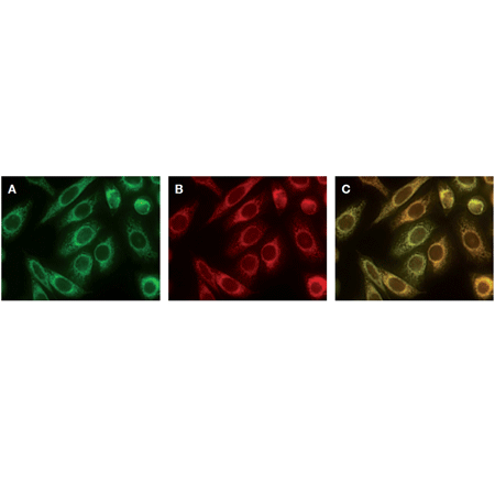
Figure 2: HeLa cells were stained with both MITO-ID® Green (A) and MITO-ID® Red (B) dyes. MITO-ID® Green dye co-localizes with the MITO-ID® Red dye, shown as yellow signal (C), demonstrating selectivity for mitochondria. Previously, MITO-ID® Red dye has been shown to co-localize with mitochondria expressing GFP.
Please mouse over
Product Details
| Applications: | Fluorescence microscopy, Fluorescent detection
|
| |
| Application Notes: | This kit has been specifically designed for use with RFP-expressing cell lines, as well as cells expressing blue or cyan fluorescent proteins (BFPs, CFPs).
One important application of MITO-ID® Green dye is in fluorescence co-localization imaging with red fluorescent protein (RFP)-tagged proteins. This is a powerful approach for determining the targeting of molecules to intracellular compartments, and for screening of associations and interactions between these molecules.
Additionally, the kit is suitable for use with live or post-fixed cells in conjunction with fluorescent probes, such as labeled antibodies, or other fluorescent conjugates displaying similar spectral properties as coumarin, phycoerythrin or cyanine-5. A nuclear counterstain (Hoechst 33342) is provided to highlight this organelle as well. |
| |
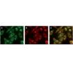
| Quality Control: | A sample from each lot of MITO-ID® Green Detection Kit is used to stain HeLa cells, using the procedures described in the user manual. The selectivity of the MITO-ID® Green dye is evident. |
| |
| Quantity: | For ENZ-51022-K500, 500 assays
For ENZ-51022-0100, 100 assays |
| |
| Shipping: | Dry Ice |
| |
| Short Term Storage: | -20°C |
| |
| Long Term Storage: | -80°C |
| |
| Contents: | MITO-ID® Green Detection Reagent
Hoechst 33342 Nuclear Stain
10x Assay Buffer
|
| |
| Technical Info/Product Notes: | The MITO-ID® Green Detection Kit is a member of the CELLESTIAL® product line, reagents and assay kits comprising fluorescent molecular probes that have been extensively benchmarked for live cell analysis applications. CELLESTIAL® reagents and kits are optimal for use in demanding cell analysis applications involving confocal microscopy, flow cytometry, microplate readers and HCS/HTS, where consistency and reproducibility are required. |
| |
| Regulatory Status: | RUO - Research Use Only |
| |
Product Literature References
GSTO1 confers drug resistance in HCT‑116 colon cancer cells through an interaction with TNFαIP3/A20: S. Paul, et al.; Int. J. Oncol.
61, 137 (2022),
Abstract;
Fumonisin B1 induces poly (ADP-ribose) (PAR) polymer-mediated cell death (parthanatos) in neuroblastoma: S. Paul, et al.; Food Chem. Toxicol.
154, 112326 (2021),
Abstract;
A mutation in the Na‐K‐2Cl cotransporter‐1 leads to changes in cellular metabolism: S. Omer, et al.; J. Cell. Physiol.
235, 7239 (2020),
Application(s): Fluorescence microscopy,
Abstract;
Full Text
Mitochondrial lipid droplet formation as a detoxification mechanism to sequester and degrade excessive urothelial membranes: Y. Liao, et al.; Mol. Biol. Cell
30, 2969 (2019),
Application(s): Fluorescence microscopy on tissue section,
Abstract;
Full Text
Constitutive regulation of mitochondrial morphology by Aurora A kinase depends on a predicted cryptic targeting sequence at the N-terminus: R. Grant, et al.; Open Biol.
8, 170272 (2018),
Application(s): Fluorescence microscopy,
Abstract;
Full Text
Therapeutically targeting mitochondrial redox signalling alleviates endothelial dysfunction in preeclampsia: C. McCarthy & L.C. Kenny; Sci. Rep.
6, 32683 (2016),
Application(s): Determination of mitochondrial mass in HUVEC ,
Abstract;
Full Text
A new and reliable method for live imaging and quantification of reactive oxygen species in Botrytis cinerea: technological advancement: R. Marschall, et al.; Fungal Genet. Biol.
71, 68 (2014),
Abstract;
General Literature References
Photoconversion of Lysotracker Red to a green fluorescent molecule.: E. C. Freundt et al.; Cell. Res.
17, 956 (2007),
Abstract;
Systematic colocalization errors between acridine orange and EGFP in astrocyte vesicular organelles.: F. Nadrigny et al.; Biophys. J.
93, 969 (2007),
Abstract;
Chloromethyl-X-rosamine (MitoTracker Red) photosensitises mitochondria and induces apoptosis in intact human cells.: T. Minamikawa et al.; J. Cell. Sci
112, 2419 (1999),
Abstract;
Chloromethyltetramethylrosamine (Mitotracker Orange) induces the mitochondrial permeability transition and inhibits respiratory complex I. Implications for the mechanism of cytochrome c release. : L. Scorrano et al.; J. Biol. Chem.
274, 24657 (1999),
Abstract;
Related Products


