- Specifically designed for use with GFP-expressing cell lines and cells expressing blue or cyan fluorescent proteins (BFPs, CFPs)
- Readily distinguishes between healthy, early apoptotic, late apoptotic and necrotic cells
- Optimized for both fluorescence microscopy and flow cytometry applications
- Suitable for death pathway analysis and drug/toxin studies
- Suitable for use with live or post-fixed cells
Enzo Life Sciences GFP CERTIFIED® Apoptosis/Necrosis Detection Kit includes all the necessary reagents for determination of early and late stages of apoptosis as well as necrosis. An Annexin V-EnzoGold (enhanced Cyanine-3) conjugate enables the detection of externalized phosphatidylserine (PS) (a marker of early apoptosis) distinct from fluorescein or GFP signals. The Necrosis Detection Reagent (Red), similarly to the red-emitting dye 7-AAD, diffuses into dead or damaged cells and binds to the DNA. Both Apoptosis Detection Reagent and Necrosis Detection Reagent are excluded from live cells with intact membranes. This kit also includes an Apoptosis Inducer (Staurosporine) for use as a positive control.
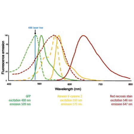
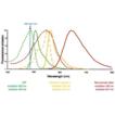
Excitation (hatched) and emission (solid) spectra for GFP, Annexin V-Cyanine 3 conjugate and Necrosis Detection Reagent (Red). All three dyes are readily excited with a 488nm laser source. The emission maxima of all fluorophores are well separated from one another.
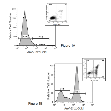
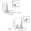
FLOW CYTOMETRY. Jurkat cells were mocked-induced with 0.2% DMSO (panel A) or induced with 2uM Staurosporine (panel B) for 4 hrs. at 37°C. After treatment, cells were incubated with a buffer containing Annexin V-Cyanine 3 and the Necrosis Detection Reagent (Red), a far red emitting DNA-intercalating dye, then analyzed by flow cytometry using a 488nm laser with fluorescence detection with FL2 (Apoptosis Detection Reagent) and FL3 (Necrosis Detection Reagent) channels. Mock-induced cells were primarily negative for apoptosis and necrosis. After a 4 hour treatment there were three populations of cells: (1) cells that were viable and not apoptotic or necrotic (Annexin V-Cyanine 3 and Necrosis Detection Reagent negative); (2) cells undergoing apoptosis (Annexin V-cyanine 3 positive and NDR negative); and (3) cells undergoing late-stage apoptosis and early necrosis (Annexin V-Cyanine 3 and Necrosis Detection Reagent positive).
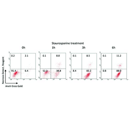
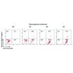
Jurkat cells stimulated with 2.5 µM Staurosporine for 0-6 h. Flow cytometry results using Jurkat suspension cells, showing early apoptotic (Annexin V-EnzoGold) and late apoptotic/necrosis (Annexin V-EnzoGold and necrosis stain).
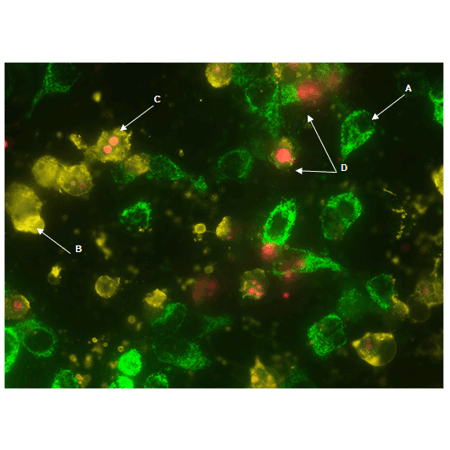
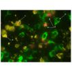
GFP-CERTIFIED Apoptosis/Necrosis Detection Kit (ENZ-51002) detects four distinct cell states. Mitochondrial GFP-expressing HeLa cells were treated with 2µM Staurosporine for 4 hours. The Apoptosis Detection Reagent (Gold) and Necrosis Detection Reagent (Red) specifically detect cell states with clear spectral separation from mitochondria-associated GFP signal. Healthy cells (A), cells undergoing apoptosis (B), cells undergoing late-stage apoptosis (C), and necrotic cells (D).
Please mouse over
Product Details
| Alternative Name: | Phosphatidylserine (PS)/DNA stain |
| |
| Applications: | Flow Cytometry, Fluorescence microscopy, Fluorescent detection
|
| |
| Use/Stability: | Store the Annexin V-EnzoGold and Binding Buffer at 4°C. Store all other reagents at -20°C. Reconstituted inducer (staurosporine) should be stored at -20°C. Refer to manual. |
| |
| Handling: | Protect from light. |
| |
| Shipping: | Blue Ice |
| |
| Short Term Storage: | +4°C |
| |
| Contents: | Apoptosis Detection Reagent (Annexin V-EnzoGold)
Necrosis Detection Reagent
Apoptosis Inducer (Staurosporine)
Binding Buffer (10X) |
| |
| Technical Info/Product Notes: | Application Note:
Image-Based Analysis of a Human Neurosphere Stem Cell Model for the Evaluation of Potential Neurotoxicants
Cited samples:
For an overview on cited samples please click here. |
| |
| Regulatory Status: | RUO - Research Use Only |
| |
Product Literature References
KRT13 is upregulated in pancreatic cancer stem-like cells and associated with radioresistance: W. Takenaka, et al.; J. Radiat. Res.
64, 284 (2023),
Abstract;
Wilforol A inhibits human glioma cell proliferation and deactivates the PI3K/AKT signaling pathway: Z. Wang, et al.; Acta Biochim. Pol.
70, 123 (2023),
Abstract;
Decreased mitochondrial function in UVA-irradiated dermal fibroblasts causes the insufficient formation of type I collagen and fibrillin-1 fibers: Y. Katsuyama, et al.; J. Dermatol. Sci.
108, 22 (2022),
Abstract;
Engineered sTRAIL-armed MSCs overcome STING deficiency to enhance the therapeutic efficacy of radiotherapy for immune checkpoint blockade: K.C.Y. Huang, et al.; Cell Death Dis.
13, 610 (2022),
Abstract;
FDA-Approved Amoxapine Effectively Promotes Macrophage Control of Mycobacteria by Inducing Autophagy: J. Wang, et al.; Microbiol. Spectr.
10, e0250922 (2022),
Abstract;
Infection of Galleria mellonella (lepidoptera) larvae with the entomopathogenic fungus Conidiobolus coronatus (entomophthorales) induces apoptosis of hemocytes and affects the concentration of eicosanoids in the hemolymph: A.K. Wronska, et al.; Front. Physiol.
12, 774086 (2022),
Application(s): Fluorescence Microscopy and Flow Cytometry of Insect Larval hemocytes,
Abstract;
Full Text
Mycobacterium tuberculosis PPE51 Inhibits Autophagy by Suppressing Toll-Like Receptor 2-Dependent Signaling: E.J. Strong, et al.; Mbio
13, e0297421 (2022),
Abstract;
Octanoic Acid - An Insecticidal Metabolite of Conidiobolus coronatus (Entomopthorales) That Affects Two Majors Antifungal Protection Systems in Galleria mellonella (Lepidoptera): Cuticular Lipids and Hemocyte: A. Kaczmarek, et al.; Int. J. Mol. Sci.
23, 5204 (2022),
Abstract;
ALL blasts drive primary mesenchymal stromal cells to increase asparagine availability during asparaginase treatment: M. Chiu, et al.; Blood Adv.
5, 5164 (2021),
Abstract;
Evofosfamide Is Effective against Pediatric Aggressive Glioma Cell Lines in Hypoxic Conditions and Potentiates the Effect of Cytotoxic Chemotherapy and Ionizing Radiations: Q. Bailleul, et al.; Cancer
13, 1804 (2021),
Abstract;
Full Text
Inhibitory role of TRIP-Br1/XIAP in necroptosis under nutrient/serum starvation: Z. Sandag, et al.; Mol. Cells
43, 236 (2020),
Abstract;
Full Text
Inhibitory role of AMP‑activated protein kinase in necroptosis of HCT116 colon cancer cells with p53 null mutation under nutrient starvation: D.T. Le, et al.; Int. J. Oncol.
54, 702 (2019),
Abstract;
Injectable and Quadruple-Functional Hydrogel as an Alternative to Intravenous Delivery for Enhanced Tumor Targeting: Z.Q. Zhang, et al.; ACS Appl. Mater. Interfaces
11, 34634 (2019),
Abstract;
Intrinsic antibacterial activity of nanoparticles made of β-cyclodextrins potentiates their effect as drug nanocarriers against tuberculosis: A. Machelart, et al.; ACS Nano
13, 3992 (2019),
Abstract;
Knockdown of TM9SF4 boosts ER stress to trigger cell death of chemoresistant breast cancer cells: Y. Zhu, et al.; Oncogene
38, 5778 (2019),
Application(s): Flow cytometry using MCF-7 cells,
Abstract;
PKM2 coordinates glycolysis with mitochondrial fusion and oxidative phosphorylation: T. Li, et al.; Protein Cell
10, 583 (2019),
Application(s): Flow cytometry using H1299 and HepG2 cells,
Abstract;
Tamoxifen acts on Trypanosoma cruzi sphingolipid pathway triggering an apoptotic death process: M. Landoni, et al.; Biochem. Biophys. Res. Commun.
516, 934 (2019),
Abstract;
Anti-leukemic efficacy of BET inhibitor in a preclinical mouse model of MLL-AF4+ infant ALL: M. Bardini, et al.; Mol. Cancer Ther.
17, 1705 (2018),
Abstract;
Pivotal role of human stearoyl-CoA desaturases (SCD1 and 5) in breast cancer progression: oleic acid-based effect of SCD1 on cell migration and a novel pro-cell survival role for SCD5: C. Angelucci, et al.; Oncotarget.
9, 24364 (2018),
Application(s): Fluorescent microscopy with MCF-7 cells,
Abstract;
Full Text
Preclinical efficacy and safety of CD19CAR cytokine-induced killer cells transfected with Sleeping Beauty transposon for the treatment of acute lymphoblastic leukemia: C.F. Magnani, et al.; Hum. Gene. Ther.
29, 605 (2018),
Abstract;
Protective effect of a newly developed fucose-deficient recombinant antithrombin against histone-induced endothelial damage: T. Iba, et al.; Int. J. Hematol.
107, 528 (2018),
Abstract;
The poly (ADP-ribose) polymerase inhibitor olaparib induces up-regulation of death receptors in primary acute myeloid leukemia blasts by NF-κB activation: I. Faraoni, et al.; Cancer Lett.
423, 127 (2018),
Application(s): Flow cytometry with primary AML cells,
Abstract;
Toxicity and phototoxicity in human ARPE-19 retinal pigment epithelium cells of dyes commonly used in retinal surgery: D. Awad, et al.; Eur. J. Ophthalmol.
28, 433 (2018),
Abstract;
Full Text
Cluster microRNAs miR‐194 and miR‐215 suppress the tumorigenicity of intestinal tumor organoids: T. Nakaoka, et al.; Cancer Sci.
108, 678 (2017),
Application(s): Flow cytometry using organoid culture of mouse intestinal tumors,
Abstract;
Full Text
Elaidic Acid, a Trans-Fatty Acid, Enhances the Metastasis of Colorectal Cancer Cells: H. Ohmori, et al.; Pathobiology
84, 144 (2017),
Abstract;
Metabolic modulation of Ewing sarcoma cells inhibits tumor growth and stem cell properties: A. Dasgupta, et al.; Oncotarget.
8, 77292 (2017),
Application(s): Flow cytometry with A673 cells,
Abstract;
Full Text
Multiple hyperthermia-mediated release of TRAIL/SPION nanocomplex from thermosensitive polymeric hydrogels for combination cancer therapy: Z.Q. Zhang, et al.; Biomaterials.
132, 16 (2017),
Application(s): Confocal microscopy with PC3 and U-87 MG cells,
Abstract;
Pro-metastatic intracellular signaling of the elaidic trans fatty acid: K. Fujii, et al.; Int. J. Oncol.
50, 85 (2017),
Abstract;
Visualization of ceramide channels in lysosomes following endogenous palmitoyl-ceramide accumulation as an initial step in the induction of necrosis: M. Yamane, et al.; Biochem. Biophys. Rep.
11, 174 (2017),
Application(s): Confocal microscopy with A549 cells,
Abstract;
Full Text
Immunotherapy of acute leukemia by chimeric antigen receptor-modified lymphocytes using an improved Sleeping Beauty transposon platform: C.F. Magnani, et al.; Oncotarget
7, 51581 (2016),
Application(s): Flow cytometry using KG-1, REH, Nalm-6, and THP-1 cells,
Abstract;
Full Text
Novel long chain fatty acid derivatives of quercetin-3-O-glucoside reduce cytotoxicity induced by cigarette smoke toxicants in human fetal lung fibroblasts: N. Sumudu, et al.; Eur. J. Pharmacol.
781, 128 (2016),
Abstract;
Phlorofucofuroeckol Improves Glutamate-Induced Neurotoxicity through Modulation of Oxidative Stress-Mediated Mitochondrial Dysfunction in PC12 Cells: J.J. Kim, et al.; PLoS One
11, e0163433 (2016),
Abstract;
Full Text
PKLR promotes colorectal cancer liver colonization through induction of glutathione synthesis: A. Nguyen, et al.; J. Clin. Invest.
126, 681 (2016),
Application(s): Assessed Apoptosis, Flow cytometry,
Abstract;
Full Text
Protective effects of β‐casofensin, a bioactive peptide from bovine β‐casein, against indomethacin‐induced intestinal lesions in rats: C. Bessette, et al.; Mol. Nutr. Food Res.
60, 823 (2016),
Abstract;
BRCA1, PARP1 and γH2AX in acute myeloid leukemia: Role as biomarkers of response to the PARP inhibitor olaparib: I. Faraoni, et al.; Biochim. Biophys. Acta
1852, 462 (2015),
Application(s): Flow cytometry using primary AML cells,
Abstract;
Growth-promoting and tumourigenic activity of c-Myc is suppressed by Hhex: V. Marfil, et al.; Oncogene
34, 3011 (2015),
Application(s): Flow cytometry using HO15.19 and TGR-1 cells,
Abstract;
High throughput proteomic analysis and a comparative review identify the nuclear chaperone, Nucleophosmin among the common set of proteins modulated in Chikungunya virus infection: R. Abraham, et al.; J. Proteomics.
120, 126 (2015),
Application(s): Flow cytometry using HEK293 cells,
Abstract;
Identification of novel osteogenic compounds by an ex-vivo sp7: luciferase zebrafish scale assay: E. de Vrieze, et al.; Blood
74, 106 (2015),
Application(s): Fluorescence microscopy using fish scales,
Abstract;
Impaired oxidative phosphorylation regulates necroptosis in human lung epithelial cells: M.J. Koo, et al.; Biochem. Biophys. Res. Commun.
464, 875 (2015),
Application(s): Apoptosis/necrosis assay,
Abstract;
Influenza virus M2 targets cystic fibrosis transmembrane conductance regulator for lysosomal degradation during viral infection: J.D. Londino, et al.; FASEB J.
29, 2712 (2015),
Application(s): Flow cytometry using HEK293 cells,
Abstract;
Full Text
Inhibition of autophagy exerts anti-colon cancer effects via apoptosis induced by p53 activation and ER stress: K. Sakitani, et al.; BMC Cancer
15, 795 (2015),
Application(s): Flow cytometric analysis of apoptosis,
Abstract;
Full Text
N-terminal functional domain of Gasdermin A3 regulates mitochondrial homeostasis via mitochondrial targeting: P.H. Lin, et al.; J. Biomed. Sci.
22, 44 (2015),
Application(s): Fluorescence microscopy using HaCaT and HEK293 cells,
Abstract;
Full Text
Novel carbocyclic curcumin analog CUR3d modulates genes involved in multiple apoptosis pathways in human hepatocellular carcinoma cells.: K.S. Bhullar, et al. ; Chem. Biol. Interact.
242, 107 (2015),
Application(s): Fluorescence microscopy,
Abstract;
NPD1-mediated stereoselective regulation of BIRC3 expression through cREL is decisive for neural cell survival: J.M. Calandria, et al.; Cell Death Differ.
22, 1363 (2015),
Application(s): Assay,
Abstract;
ROS-induced oxidative stress and apoptosis-like event directly affect the cell viability of cryopreserved embryogenic callus in Agapanthus praecox: D. Zhang, et al.; Plant Cell Rep.
34, 1499 (2015),
Abstract;
The Adhesion GPCR CD97/ADGRE5 inhibits apoptosis: C.C. Hsiao, et al.; Int. J. Biochem. Cell. Biol.
65, 197 (2015),
Application(s): Flow cytometry using HT1080 cells,
Abstract;
The effect of plasma-derived activated protein C on leukocyte cell-death and vascular endothelial damage: T. Iba, et al.; Thromb. Res
135, 963 (2015),
Application(s): Fluorescence Microscopy using leukocytes,
Abstract;
Accumulation of cytosolic calcium induces necroptotic cell death in human neuroblastoma: M. Nomura, et al.; Cancer Res.
74, 1056 (2014),
Abstract;
Apoptotic and inhibitory effects on cell proliferation of hepatocellular carcinoma HepG2 cells by methanol leaf extract of Costus speciosus: S.V. Nair, et al.; Biomed. Res. Int.
2014, Article ID 637098 (2014),
Application(s): Measurement by flow cytometry,
Abstract;
Full Text
Combination of antithrombin and recombinant thrombomodulin modulates neutrophil cell-death and decreases circulating DAMPs levels in endotoxemic rats: T. Iba, et al.; Throm. Res.
134, 169 (2014),
Application(s): Detection Kit using live cells,
Abstract;
Fatty acid esters of ohloridzin induce apoptosis of human liver cancer cells through altered gene expression: S.V. Nair, et al.; PLoS One
9, e107149 (2014),
Application(s): Fluorescence microscopy of HepG2 cells,
Abstract;
Full Text
Flavonoid-enriched apple fraction AF4 induces cell cycle arrest, DNA topoisomerase II inhibition, and apoptosis in human liver cancer HepG2 cells: S. Sudan, et al.; Nutr. Cancer
66, 1237 (2014),
Abstract;
Quercetin-3-O-glucoside induces human DNA topoisomerase II inhibition, cell cycle arrest and apoptosis in hepatocellular carcinoma cells: S. Sudan, et al.; Anticancer Res.
34, 1691 (2014),
Abstract;
A light-activated NO donor attenuates anchorage independent growth of cancer cells: Important role of a cross talk between NO and other reactive oxygen species: S. Sen, et al.; Arch. Biochem. Biophys.
540, 33 (2013),
Application(s): Detection Kit using live cells,
Abstract;
Alu Elements in ANRIL Non-Coding RNA at Chromosome 9p21 Modulate Atherogenic Cell Functions through Trans-Regulation of Gene Networks: L.M. Holdt, et al.; PLoS Genet.
9, e1003588 (2013),
Application(s): Detection Kit using live cells,
Abstract;
Full Text
Analysis of protein translocation into the endoplasmic reticulum of human cells: J. Dudek, et al.; Methods Mol. Biol.
1033, 285 (2013),
Abstract;
Characterizing virulence-specific perturbations in the mitochondrial function of macrophages infected with Mycobacterium tuberculosis: S. Jamwal, et al.; Sci. Rep.
3, 1328 (2013),
Abstract;
Full Text
Evaluation of anticancer effects and enhanced doxorubicin cytotoxicity of xanthine derivatives using canine hemangiosarcoma cell lines: T. Motegi, et al.; Res. Vet. Sci.
95, 600 (2013),
Application(s): Fluorescence microscopy using canine HAS cells,
Abstract;
Genista sessilifolia DC. extracts induce apoptosis across a range of cancer cell lines: P. Bontempo, et al.; Cell Prolif.
46, 183 (2013),
Application(s): Detection Kit using live cells,
Abstract;
High mobility group box 1 released from necrotic cells enhances regrowth and metastasis of cancer cells that have survived chemotherapy: Y. Luo, et al.; Eur. J. Cancer.
49, 741 (2013),
Application(s): Fluorescence microscopy using CT26 cells,
Abstract;
Influence of gefitinib and erlotinib on apoptosis and c-MYC expression in H23 lung cancer cells: M. Suenaga, et al.; Anticancer Res.
33, 1547 (2013),
Abstract;
Comparison of different suicide-gene strategies for the safety improvement of genetically manipulated T cells: V. Marin, et al.; Hum. Gene Ther. Methods
23, 376 (2012),
Application(s): Flow cytometry using EBV-CTL and 293 T cells,
Abstract;
Full Text
Maintenance of higher H2O2 levels, and its mechanism of action to induce growth in breast cancer cells: Important roles of bioactive catalase and PP2A: S. Sen, et al.; Free Radic. Biol. Med.
53, 1541 (2012),
Application(s): Detection Kit using live cells,
Abstract;
Mesenchymal stem cells from Shwachman–Diamond syndrome patients display normal functions and do not contribute to hematological defects: V. André, et al.; Blood Cancer J.
2, e94 (2012),
Application(s): Flow cytometry using human neutrophils,
Abstract;
Full Text
Methionine excess in diet induces acute lethal hepatitis in mice lacking cystathionine γ-lyase, an animal model of cystathioninuria: H. Yamada, et al.; Free Radic. Biol. Med.
52, 1716 (2012),
Application(s): Fluorescence microscopy using mouse hepatocytes,
Abstract;
Molecular mechanism of interleukin-2-induced mucosal homeostasis : J. Mishra, et al.; Am. J. Physiol. Cell Physiol.
302, C735 (2012),
Application(s): Detection Kit using live cells,
Abstract;
Full Text
Serratia marcescens induces apoptotic cell death in host immune cells via a lipopolysaccharide- and flagella-dependent mechanism: K. Ishii, et al.; J. Biol. Chem.
287, 36582 (2012),
Application(s): Apoptosis detected in Silkworm larvae,
Abstract;
Two mutations impair the stability and function of ubiquitin-activating enzyme (E1): T. Lao, et al.; J. Cell. Physiol.
227, 1561 (2012),
Application(s): Viability and induction of cell death observed by confocal microscopy,
Abstract;
Full Text
Critical roles of Cold Shock Domain Protein A as an endogenous angiogenesis inhibitor in skeletal muscle: Y. Saito, et al.; Antioxid. Redox. Signal.
15, 2109 (2011),
Abstract;
Cell Surface Externalization of Annexin A1 as a Failsafe Mechanism Preventing Inflammatory Responses during Secondary Necrosis: K.E. Blume, et al.; J. Immunol.
183, 8138 (2009),
Abstract;
Full Text
Supplemental Information for: Cell Surface Externalization of Annexin A1 as a Failsafe Mechanism Preventing Inflammatory Responses during Secondary Necrosis: K.E. Blume, et al.; J. Immunol.
183, (2009), (Supplemental Information),
Full Text
Related Products


